
May 2022 – Presented by Dr. Melissa Ha (Mentored by Dr. Han Sung Lee)
The patient is a 66-year-old female referred for consultation regarding an incidentally found 4 cm renal mass of the right kidney as well as a 4 cm soft tissue pelvic mass at the bifurcation of the right iliac vessels on CT imaging. She had initially presented for a routine health check with elevated LFTs. The patient underwent surgical management. Open abdominal surgery revealed an appendiceal mass that correlated with the pelvic mass on imaging. The patient received a total nephrectomy and an appendectomy. The renal mass was diagnosed to be clear cell renal cell carcinoma.
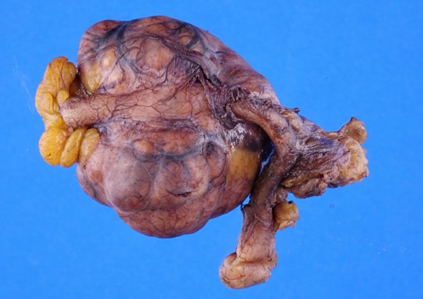
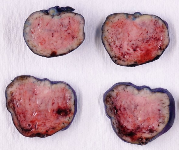
Gross examination of the appendectomy specimen revealed a purple tan bulging nodular mass measuring 4.5 x 4.5 x 3.3 cm located at the distal appendix and is entirely encased within the mesoappendix. The mass focally obliterates the entire cross-section of the appendix and has homogeneous pink tan cut surfaces with areas of hemorrhage. No necrosis was noted.
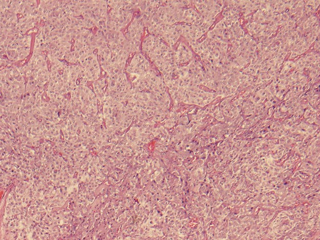
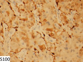
Histologic evaluation shows clustered and nested growth pattern separated by prominent fibrovascular stroma. Neoplastic cells feature fine granular basophilic cytoplasm and salt and pepper nuclei. Immunohistochemical studies show tumor cells stain GATA3 positive, AE1/AE3 negative with low proliferation index (Ki67 less than 1%). S100 stains scattered cells present around tumor nests.
Click to enlarge the images!
 Meet our Residency Program Director