3D models provide road map to cloacal repairs
This video is best viewed in Chrome, Firefox or Safari.
(SACRAMENTO) — Cloacal repair is a delicate surgical procedure that many pediatric surgeons will never encounter in their lifetime.
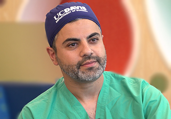
But UC Davis pediatric surgeon Payam Saadai specializes in this type of surgery. He is committed to helping children born with a cloaca to have the best possible future.
One in 50,000 girls are born with a cloaca, which is a rare, congenital malformation in which the genital tract, the urinary tract and the colorectal or intestinal tract end together in one channel, instead of being three separate structures.
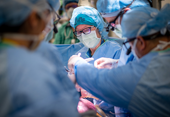
The surgery to reconstruct those three separate structures takes place in a single surgery and is a collaborative effort by a team of experienced pediatric surgeons, pediatric urologists, interventional radiologists, gynecologists and other specialists.

Payam Saadai
“No two cases are the same,” said Saadai, who performs four to five cloacal repairs on infants each year at UC Davis Children’s Hospital in close collaboration with UC Davis pediatric urologist Eric Kurzrock. He waits until babies are nine months to a year old before performing this life-changing surgery. “Everyone is a little different. The way the structures connect is different. Some connect very high up in the belly. Some of them connect really low down. Sometimes, there will be abnormalities related to the urological tract or the gynecological tract that can confuse things. If you go into surgery without a good road map, you could run into complications.”
Three-dimensional printed models have helped the surgical teams plan for this complex surgery. These models provide a road map of sorts that is making all the difference.
A complex anatomy in 3D
Each cloacal model is made at the 3D PrintViz Lab on the UC Davis Health campus, based on two-dimensional scans of the patient’s anatomy from radiology and 3D imaging known as cloacagrams.
Osama Raslan, associate professor of radiology at UC Davis Health and co-founder of the 3D PrintViz Lab, has partnered with Saadai to provide the custom models needed to help the surgical team visualize the complexity of the anatomy, plan for the surgery and anticipate complications.
“Models do what two-dimensional scans can’t do. 3D takes away the guesswork,” Raslan said. “You can hold the 3D model and change the positioning in your hands. You can see different perspectives in a way that you can’t with X-rays.”
The benefits of 3D models over 2D scans
The 3D lab uses FDA-approved software to create the models. Models are printed on site.
By inputting 2D images and scans into the software, Raslan can separate the organs or systems that the surgeons are interested in seeing. He can also color code them to highlight different organs or body systems.
Some advantages of the 3D model over 2D scans:
- Models are portable and replicate the exact size of the patient’s organs and anatomy.
- Models can be made of sterile materials and brought into the operating room. Surgeons can refer to the model during surgery.
- Models can be assembled or disassembled, allowing clinicians to take apart and reconstruct the anatomy.
- Surgeons can move the model around and flip it, providing different perspectives from all sides.
- Models can help parents understand what will be taking place during surgery.
- Models can be a teaching tool for medical residents and fellows so they can see the complex anatomy and the surgical planning required.
Raslan is grateful that his lab can use this technology to help surgeons be successful, especially in complex cases like cloacal repair.
“This technology can drastically change a patient’s life for the better. That’s the most important reason why we’re doing this,” Raslan said.
Saadai remembers a time when he didn’t have access to 3D models to complete cloacal repairs. He said that the outcome was still the same. It just took longer and was more challenging to get the same results. Thankfully he doesn’t have to work in that environment any longer.
“If we didn’t have the 3D modeling here at UC Davis, it would be more difficult to achieve the same results. Most centers don’t have this capability. Most surgeons who do this cloacal repair don’t have access to 3D models. We’re in a very unique situation here at UC Davis. I feel very lucky,” Saadai said.


