Residency Program - Case of the Month
July 2012 - Presented by Mahan Matin, M.D.
Clinical history:
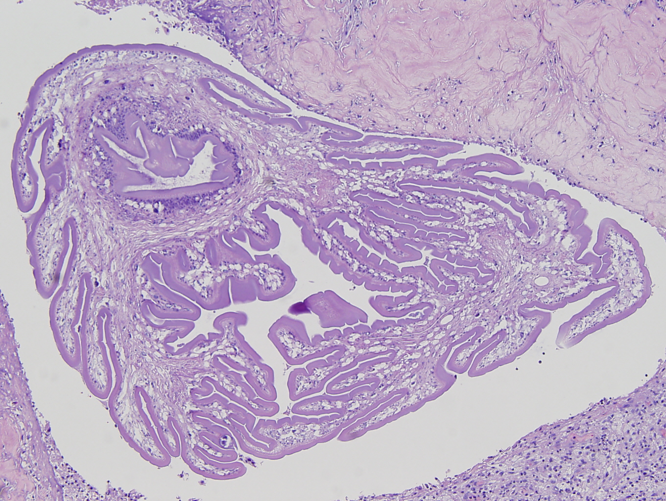
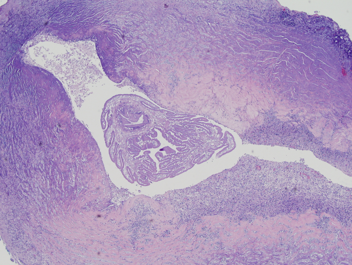
The patient is an 11-year-old girl with no significant past medical history who presented with a headache located behind the right eye, with radiation to bilateral temples. She complained of blurry vision and seeing sparkles in the right eye. She had emesis but no nausea. There was no clear history of seizure but she had difficulty understanding language, with slight weakness in the right upper extremity. MRI showed a lesion at the periphery of the right frontal cortex with surrounding edema, a rim of enhancement, and two hypointense central foci. In the operating room, an organized and firm tissue with small pockets of pus was identified underneath the dura. The tissue was easy to separate from the surrounding brain and the lesion was completely excised.
Gross description:
Received in formalin was a 1.1 x 0.7 x 0.4 cm pink-tan soft tissue. The outer surface was inked blue. The specimen was sectioned and entirely submitted in one cassette.
Microscopic images:


 Meet our Residency Program Director
