
Residency Program - Case of the Month
January 2013 - Presented by Brian Gorospe, M.D.
Clinical history:
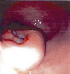
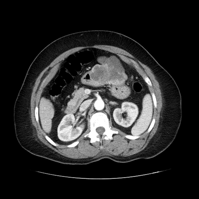
A 63-year-old female with a past medical history of gastrointestinal bleeding from a gastric ulcer and H. pylori infection presented with new complaints of melena, progressive weakness, fatigue and shortness of breath. Labs were consistent with iron deficiency anemia and a hemoglobin of 3.2 g/dL. An upper endoscopy revealed a large, ulcerated gastric mass between the incisura and antrum, concerning for malignancy (Figure 1). Abdominal CT scan demonstrated a 9.0 x 6.0 x 5.5 cm multilobulated enhancing mass involving the body and antrum, extending beyond the borders of the stomach (Figure 2).
Images:
| Figure 1 | Figure 2 | |
 |
 |
Endoscopic biopsy:
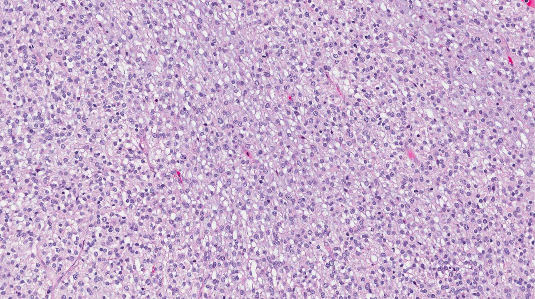
An endoscopic biopsy of the gastric mass was performed and included fragments of benign gastric mucosa with ulceration, as well as a small fragment of a poorly differentiated epithelioid neoplasm (Figure 3).
| Figure 3 |
 |
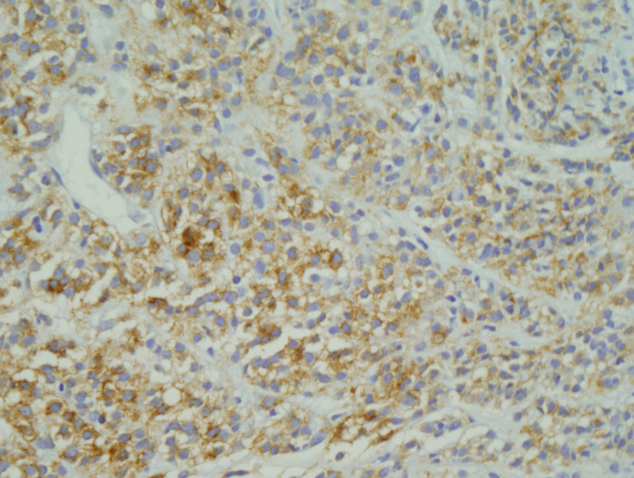
Immunohistochemical stains:
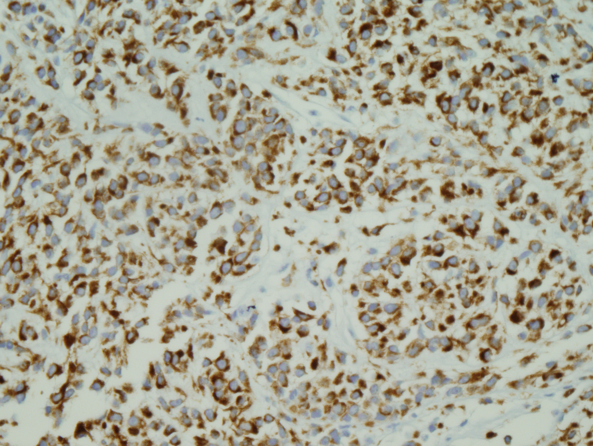
| AE1/AE3: | positive (figure 4) |
| CD117: | positive (figure 5) |
| CD45: | negative |
| S100: | negative |
| Desmin: | negative |
| Figure 4 | Figure 5 | |
 |
 |
 Meet our Residency Program Director