
Facilities
Sacramento
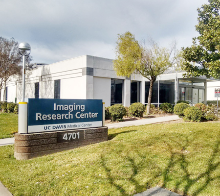
The IRC's primary site is a 13,000 sq. ft. building on the UC Davis Health campus in Sacramento. It houses a research-dedicated whole-body 3T Siemens Prisma Fit MRI system, offices and shared workspace for investigators, technical staff, and trainees, and general imaging research support equipment.
A wide range of imaging studies is performed at the IRC. These include basic science and clinical research on the structure and function of the nervous system (including perceptual, motor, and cognitive function, studied with real-time functional imaging), and research on systemic physiology and morphology in health and disease.
Siemens Prisma Fit Scanner
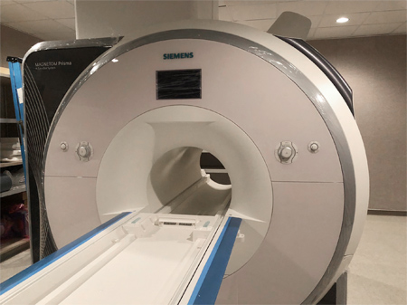
The IRC operates a 3T Prisma Fit whole-body MRI system (Siemens Healthineers, Erlangen, Germany), which is fully equipped for advanced brain and body imaging. The system has a short-bore (2 m) magnet, a fast gradient system that provides high-speed structural and functional imaging, and a 64-channel data acquisition system for parallel imaging.
The Prisma Fit combines a maximum gradient amplitude of 80 mT/m with a 200 T/m/s slew rate, which delivers higher SNR throughout the body. Powerful gradients are critical for fast imaging techniques such as Echo Planar Imaging (EPI). EPI is the modality of choice for both functional MRI (fMRI) and diffusion-weighted imaging (DWI), which UC Davis investigators use extensively.
The Prisma Fit uses a parallel transmit system with two independent coils. Varying the relative phase between the two transmit coil elements homogenizes the B1 field. This is essential for signal uniformity across the field of view, and it also speeds up scans limited by the specific absorption rate.
The improved radio frequency (RF) transmit and receive system uses digital input and output and carries the RF signal over fiber-optic cables. This eliminates electromagnetic crosstalk between RF channels and interference from spurious RF sources, improving the signal-to-noise ratio (SNR). The higher spatial and temporal SNR shortens total exam time, so studies involving children or patients with anxiety can get higher-quality data in less time.
Dedicated force compensation on each gradient axis keeps vibration and acoustic noise low. High-performance cooling for each axis allows a full duty cycle over long measurements with outstanding stability. As a result, the Prisma Fit's gradients produce significantly less noise than previous systems, which is expected to increase both participant comfort and the percentage of participants who complete their scanning sessions.
Three multi-channel RF head coils are available:
- a Siemens 64-channel brain coil, which takes full advantage of the 64 receiver channels and provides the highest signal-to-noise ratio;
- a 32-channel coil, which provides continuity with ongoing studies acquired on the previous TIM Trio system;
- a 16-channel coil, which provides excellent SNR while leaving more space around the head for headphones and goggles, for enhanced auditory and visual presentation and simultaneous EEG/fMRI studies.
A range of coils for other body regions is also available. The system hardware, RF coils, and software support acceleration techniques such as parallel imaging and simultaneous multi-slice. Current software packages include diffusion tensor imaging, 2D arterial spin labeling perfusion imaging, susceptibility-weighted imaging, BOLD resting-state fMRI, single-voxel spectroscopy, 2D and 3D chemical shift imaging, and multi-nuclear spectroscopy. The system software is scheduled to be upgraded to syngo MR XA60 on November 30, 2026, which will make a range of new pulse sequences available.
CO2 Challenge Setup
The IRC houses a comprehensive setup for CO2 challenges, including an MR-compatible, computer-controlled gas delivery system, specialized breathing circuits, and continuous physiological monitoring. This infrastructure supports advanced cardiovascular and neurovascular imaging with high standards of safety and reproducibility.
Shared Use TMS Laboratory
The Shared Use TMS Laboratory provides a dedicated environment for noninvasive transcranial magnetic stimulation (TMS), to support sham stimulation procedures in human participants. It is designed to closely emulate the conditions of a clinical TMS suite while preventing effective cortical stimulation during control conditions.
The laboratory is equipped with a Magstim Super Rapid TMS system with a figure-8 coil, air-cooled by a vacuum-powered fan and used exclusively for sham stimulation. A neuronavigation system from Rogue Industries (Standish, ME, USA) positions the sham coil over the intended stimulation site, based on each participant's structural or functional MRI data. This lets sham procedures mirror the spatial targeting and participant experience of active stimulation in a clinical facility.
The TMS laboratory is available to all IRC users. To schedule it, please contact the IRC.