Heart and VascularJuly 27, 2021
UC Davis Health fixes baby’s heart defect using echocardiography-only strategy
Patent ductus arteriosus closure successfully completed without contrast
(SACRAMENTO)
Cedric Taylor was born at 25 weeks. Like many premature babies, he was born with a heart defect called patent ductus arteriosus (PDA).
PDA is a condition in which the ductus arteriosus, a blood vessel in the heart that is open before birth to divert blood from the lungs to the body, does not close spontaneously after birth.
Of the four million annual births in the U.S., about 1.5% (60,000) of babies are born severely premature with very low birth weight (less than 3.3 pounds). Twenty percent (12,000) of these have a significant PDA.
Without treatment for PDA, excessive blood flow to the lungs can increase pressure in the pulmonary arteries. It can lead to significant medical problems, including acute kidney injury, brain bleeds, ischemic injury of the intestines, pulmonary vascular disease and heart failure. With excessive blood to the lungs, the respiratory system deteriorates and babies experience a decreased tolerance to feeding, resulting in poor weight gain.
For more than three years, UC Davis Health has been successfully performing PDA closures in the cardiac catheterization lab on babies as small as 600 grams or 1.3 pounds.
By implanting an Amplatzer Piccolo Occluder medical device, the world’s smallest FDA-approved PDA device, through a catheter in the groin, doctors can close the ductus arteriosus without surgery. Patients can see dramatic improvements in lung function immediately as shown on their chest X-rays.
In Taylor’s case, the procedure posed challenges. Taylor weighed 930 grams (2.05 lbs.) at the time of the procedure. His kidneys weren’t functioning well, caused by poor blood flow due to the large PDA. The use of contrast (dye) needed to take pictures (angiograms) during the procedure was risky.
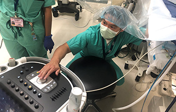
The procedure was guided by echo images from Jay Yeh.
“In order to close the PDA, we have to give him contrast to be able to take X-ray pictures before and after the occlusion and see the anatomy clearly. We need the angiograms to make sure we are placing the Piccolo medical device into the proper position. But the contrast used can potentially cause further harm to the kidneys that are already not working well. It was a difficult catch-22 situation,” said interventional cardiologist and chief of pediatric cardiology Frank Ing, who would perform the PDA closure.
It was too risky. So, the team came up with a new strategy: a PDA closure guided only by heart ultrasound, better known as echocardiography (echo) without contrast or angiograms.
A new way to PDA
Is it possible to solely use echo to help guide the placement of the Amplatzer Piccolo Occluder device without traditional angiography? Could echo replace angiography, which uses contrast and X-rays to show the anatomy of the PDA?
Ing discussed the novel strategy with fellow pediatric cardiologist and medical director of the pediatric echocardiography lab Jay Yeh.
“It’s not always easy to see blood vessels by echo,” Yeh said. “But Taylor had an echo a couple of days prior. I felt pretty confident that I could see the position of the device in the PDA and ensure that the device would not cause flow obstruction to nearby blood vessels. I felt that I could guide Dr. Ing to the proper position for the device implant, based on what I was seeing in the echo.”
They agreed to try it.
During the cardiac catheterization procedure, Ing passed a thin catheter into a blood vessel in Taylor’s groin and moved it up into the heart, only guided by echo images provided by Yeh, to locate the PDA. Inserting the Amplatzer Piccolo Occluder medical device into the catheter and into the PDA, Dr. Ing was able to successfully release the device to block the abnormal blood flow of the PDA.
In less than two hours, the procedure was complete.
Taylor’s kidney function significantly improved the next morning. A post-PDA echo revealed that no residual PDA remained. The procedure was an overwhelming success.
“I am aware of only a few interventional cardiologists in the country who have done this,” Ing said. “I am proud that we were able to help this baby using a team approach.”
Cedric Taylor’s mom Candra is just thankful that this procedure has made a big difference to her son’s health overnight.
“I appreciate Dr. Ing for taking the risk and making the best decision for Cedric, due to certain circumstances. Dr. Ing did great with reassuring that he only had confidence that the procedure would be successful,” said Taylor.
Cedric will not require any additional PDA procedures. The device will remain in place for Cedric’s lifetime.
“Baby Cedric clinically is doing much better. We are happy and growing. Only ups and gains from here!” Taylor said.