Dogs get head and neck cancers, too
Clinical trial in canine companions may help humans with cancer
When Sarah Lindley found a lump near her dog Bucky’s tooth, she didn’t think it was a problem. The lively husky mix, which she and her partner, Tom Yuzvinsky, consider part of the family, didn’t appear to be in pain. Still, she scheduled an appointment with her local veterinarian on the Central Coast.
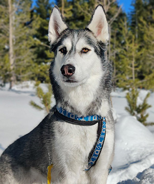
“At first we thought something was stuck in his gums and he might lose a tooth,” Lindley said. “Then the biopsy came back as cancerous.”
To get treatment for Bucky as quickly as possible, the couple turned to the UC Davis School of Veterinary Medicine. He was diagnosed with squamous cell carcinoma, the second-most common oral cancer in dogs, according to Stephanie Goldschmidt, an assistant professor caring for companion pets at the veterinary school’s Veterinary Medical Teaching Hospital.
Before removing Bucky’s cancerous tumor surgically, Goldschmidt asked to enroll him in a clinical trial funded by UC Davis Comprehensive Cancer Center. Cancer in dogs is similar to cancer in people. Bucky and other dogs are helping scientists advance cancer cell detection, with the potential to improve oral surgery outcomes for canines and their human companions.
Cancer treatments in pets that can help people
It’s called comparative oncology and it holds the promise of transforming cancer treatments for pets as well as people. Goldschmidt is part of the new Head and Neck Malignancies Innovation Group at the cancer center. The clinical trial in which Bucky is enrolled exemplifies the collaborative research the group is doing.
“Squamous cell carcinoma is the most common oral cancer in humans, and the mutational profile for malignant cells in canines and humans is quite similar,” explained Goldschmidt, the project’s lead researcher. “We think that dogs provide an excellent model for [studying] the disease.”
Squamous cell carcinoma recurs close to 30% of the time in dogs. Recurrence is also high in humans. In fact, surgeries for this type of cancer have the third-highest positive margins, indicating that malignant cells remain after the tumor is removed. As standard practice, surgeons remove a border of healthy tissue around tumors to catch stray cancer cells and thereby prevent cancer recurrence.
Minimizing side effects from surgery
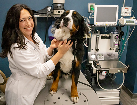
Surgeons observe a fine balance between removing enough tissue to prevent cancer regrowth and preserving quality of life. In Goldschmidt’s experiences, dog tumors tend to be more advanced by the time they are discovered and often require removal of bone tissue. Although reconstructive surgery isn’t always an option, she and her team are adept at correcting postoperative defects, as she did for Bucky after removing the front part of his lower jaw.
In both dogs and humans, surgery can impair various functions. Tumor removal in the head and neck region can significantly alter vocal capabilities, swallowing and appearance, explained Andrew Birkeland, who is an associate professor in the Department of Otolaryngology Head and Neck Surgery and one of the co-directors of the Head and Neck Malignancies Innovation Group.
Goldschmidt’s research is designed to pinpoint tumor margins with greater accuracy and help surgeons spare more healthy tissue. “Taking the most conservative route with tissue removal while preventing cancer spread is always the best option,” she said.
Auto-fluorescence lights the way
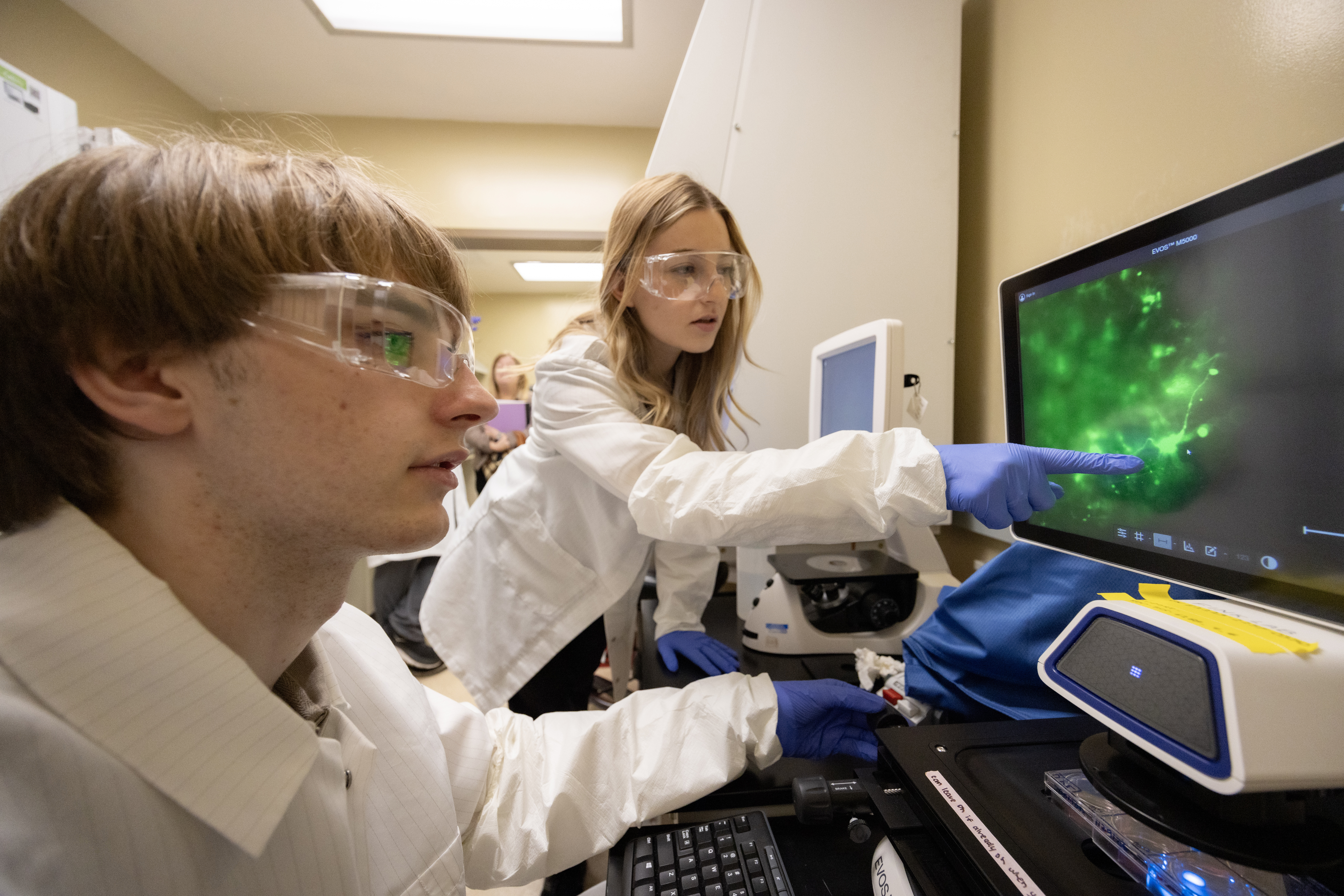
The members of the oncology clinical trial team, which includes veterinary pathologists and oncologists as well as biomedical engineers, are evaluating a cancer-detection technique using the oral fluorophore 5-ALA. It emits light of a certain wavelength when stimulated by lasers.
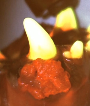
Oral tissues have their own luminosity, called auto-fluorescence, that researchers can see using spectral imaging. In cancer cells, the spectral properties are shifted. In the trial that includes Bucky, the researchers are determining if 5-ALA, an amino acid commonly found in mammal cells, would make changes in fluorescence properties between normal and cancer tissue more pronounced than focusing on auto-fluorescence alone.
Before surgery, Goldschmidt gives canine participants a pill containing 5-ALA, an amino acid that breaks down into the compound protoporphyrin IX. It tends to accumulate in cancer cells. Using a fluorescence lifetime imaging device developed and built in Biomedical Engineering Professor Laura Marcu’s lab, the team screens for protoporphyrin IX and auto-fluorescence spectral properties and compares those measurements to tumor margins after surgical removal.
If the trial succeeds, the team will progress to 5-ALA research in human head and neck patients. They are currently analyzing their data and determining the potential to train computers to track cancer cells using spectral images.
Marcu and colleagues in the Department of Neurological Surgery’s UC Davis National Center for Interventional Biophotonic Technologies are also using Interventional Fluorescence Lifetime Imaging to identify tumor margin, histology, and grade of malignant brain tumors.
Hope for canine cancer patients
Bucky’s family members were excited to assist with the trial. “It wasn’t a difficult decision. Goldschmidt and the rest of the UC Davis team were so obviously committed to their work. It felt good to give back,” Yuzvinsky said.
After facial reconstruction, Bucky was eating on his own and holding a ball in his mouth. The couple looks forward to getting back to their normal lives post-surgery, like skijoring, when Bucky and the other family dog enjoy pulling them on skis.
“Basically, he loves his humans,” said Lindley. “He just loves his pack.”
UC Davis Comprehensive Cancer Center
UC Davis Comprehensive Cancer Center is the only National Cancer Institute-designated center serving the Central Valley and inland Northern California, a region of more than 6 million people. Its specialists provide compassionate, comprehensive care for more than 100,000 adults and children every year and access to more than 200 active clinical trials at any given time. Its innovative research program engages more than 240 scientists at UC Davis who work collaboratively to advance discovery of new tools to diagnose and treat cancer. Patients have access to leading-edge care, including immunotherapy and other targeted treatments. Its Office of Community Outreach and Engagement addresses disparities in cancer outcomes across diverse populations, and the cancer center provides comprehensive education and workforce development programs for the next generation of clinicians and scientists. For more information, visit cancer.ucdavis.edu.


