
Residency Program - Case of the Month
August 2012 - Presented by Aram Millstein, M.D.
Clinical history:
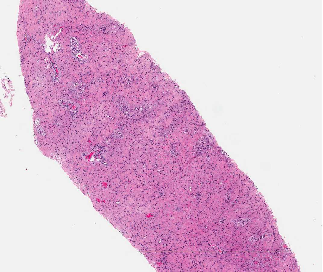
The patient is a 44 year old pravida 2, para 2 woman with a seven-year history of a pedunculated vulvar mass. She initially noticed a pruritic mass-like lesion with occasional pain. During her last pregnancy five years ago, an attempt to drain this mass was made but without any output from it. A recent ultrasound showed a 7-cm mass with high vascularity. Physical exam showed a left-sided mass coming off the left labia and extending to the posterior fourchette. Medially it extends up to the vagina but not into it. The mass appears sessile and is completely covered by intact skin. A core biopsy of the mass was performed. The histology is shown below.
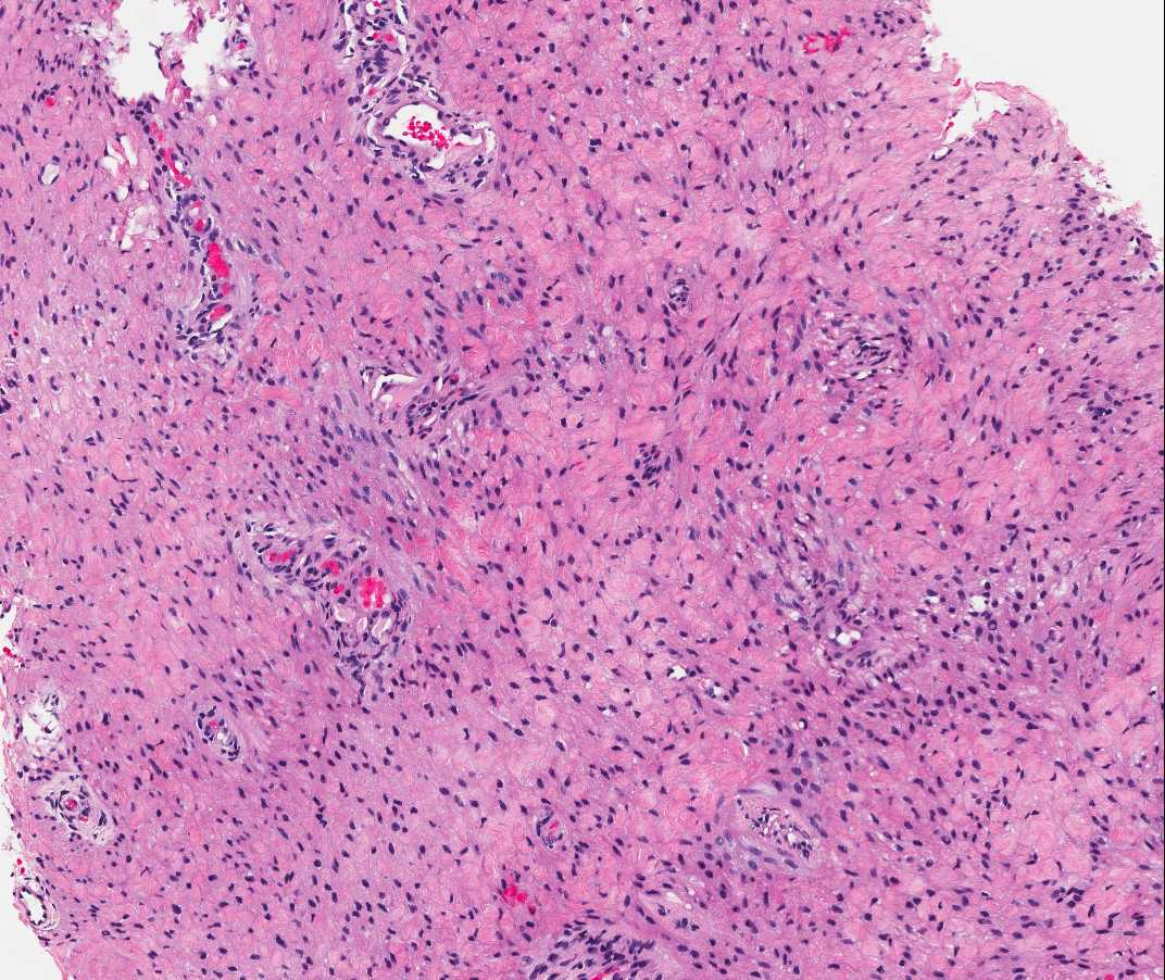
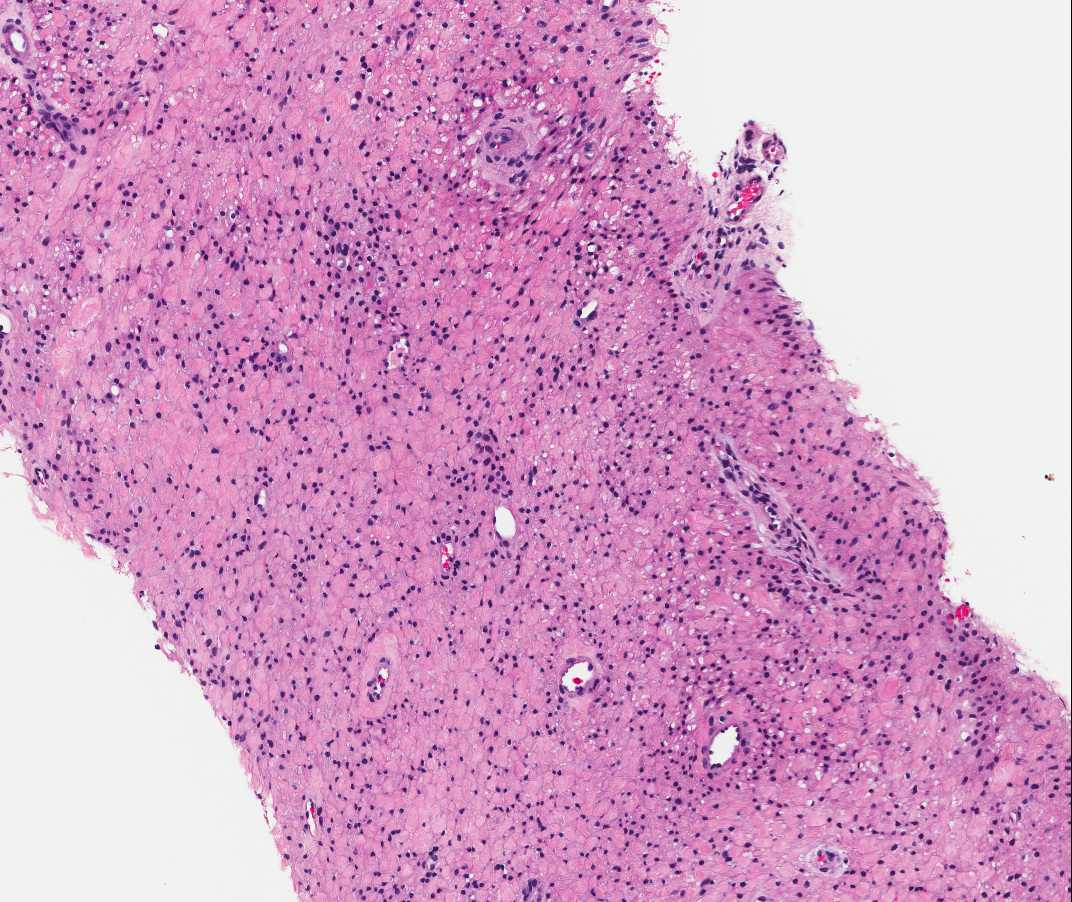
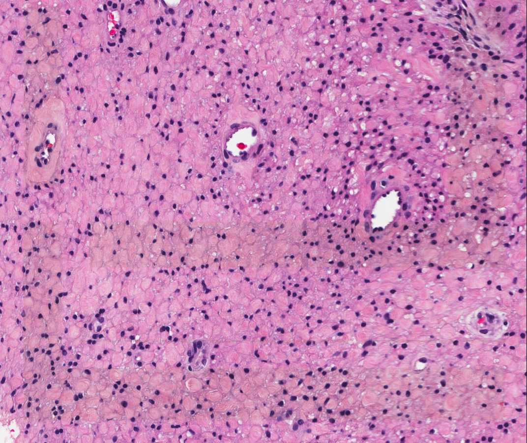
Microscopic images:
 |
 |
 |
 |
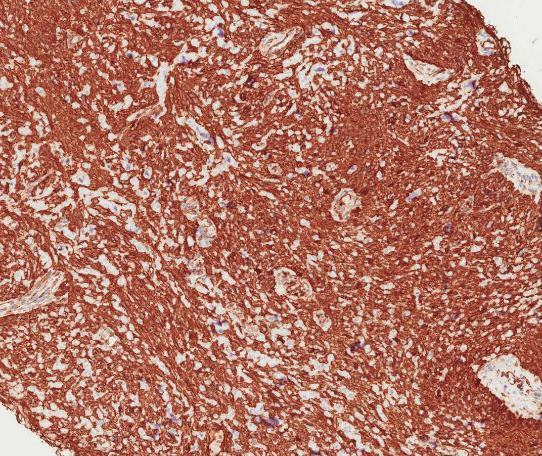
Immunohistochemistry:
| Smooth muscle actin | CD34 | Projesterone receptor | ||||
 |
 |
 |
||||
| Estrogen receptor | S100 | |||||
 |
 |
 Meet our Residency Program Director