Residency Program - Case of the Month
January 2015 - Presented by Dr. Nicholas Coley & Dr. Gloria Lewis, Mather Veteran Affairs
Clinical History & Histology
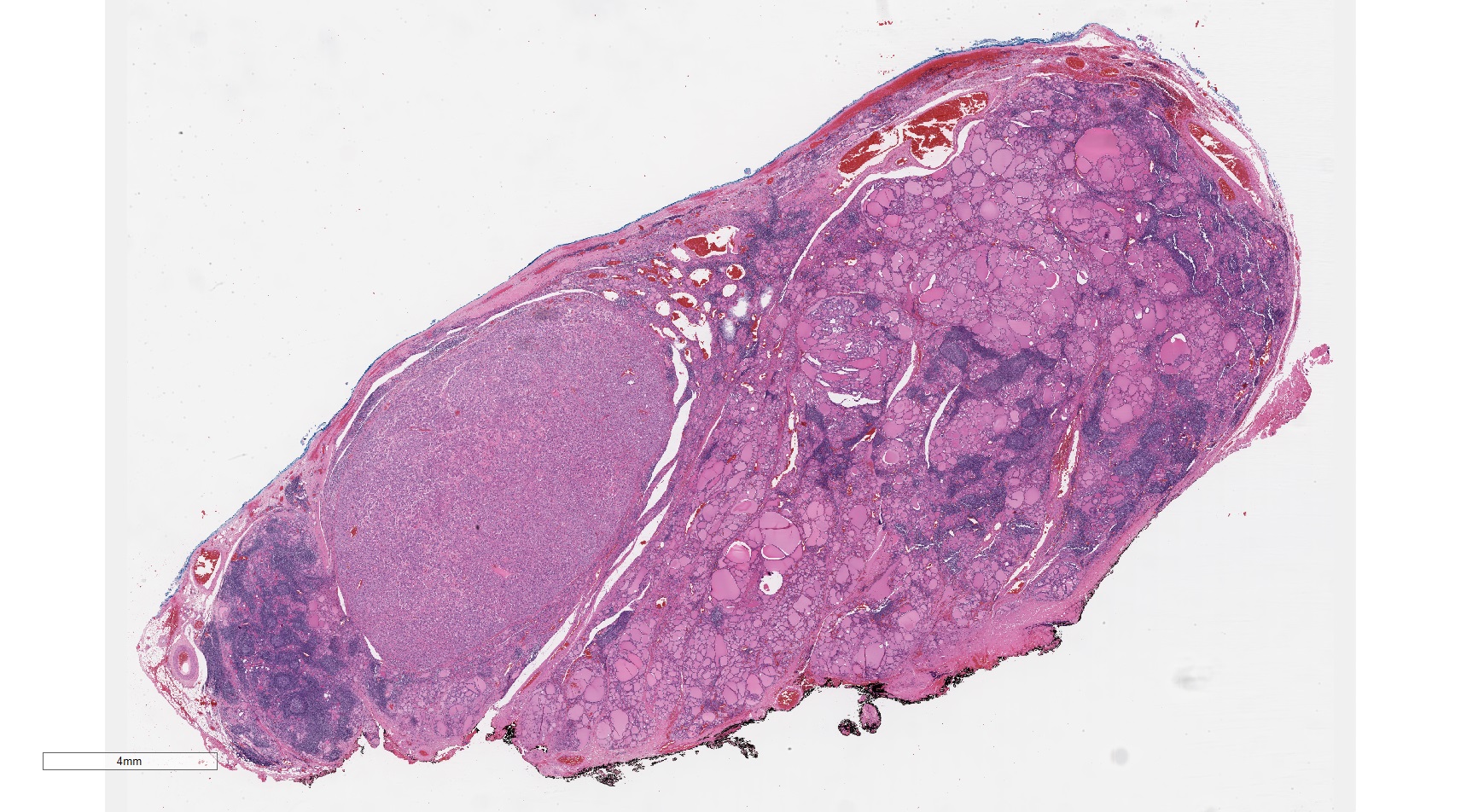
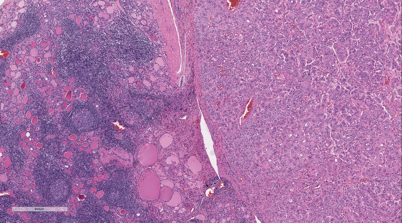
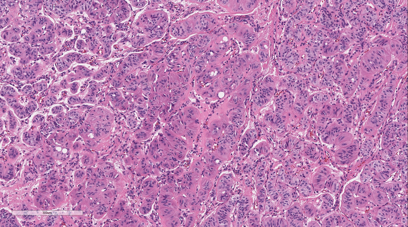
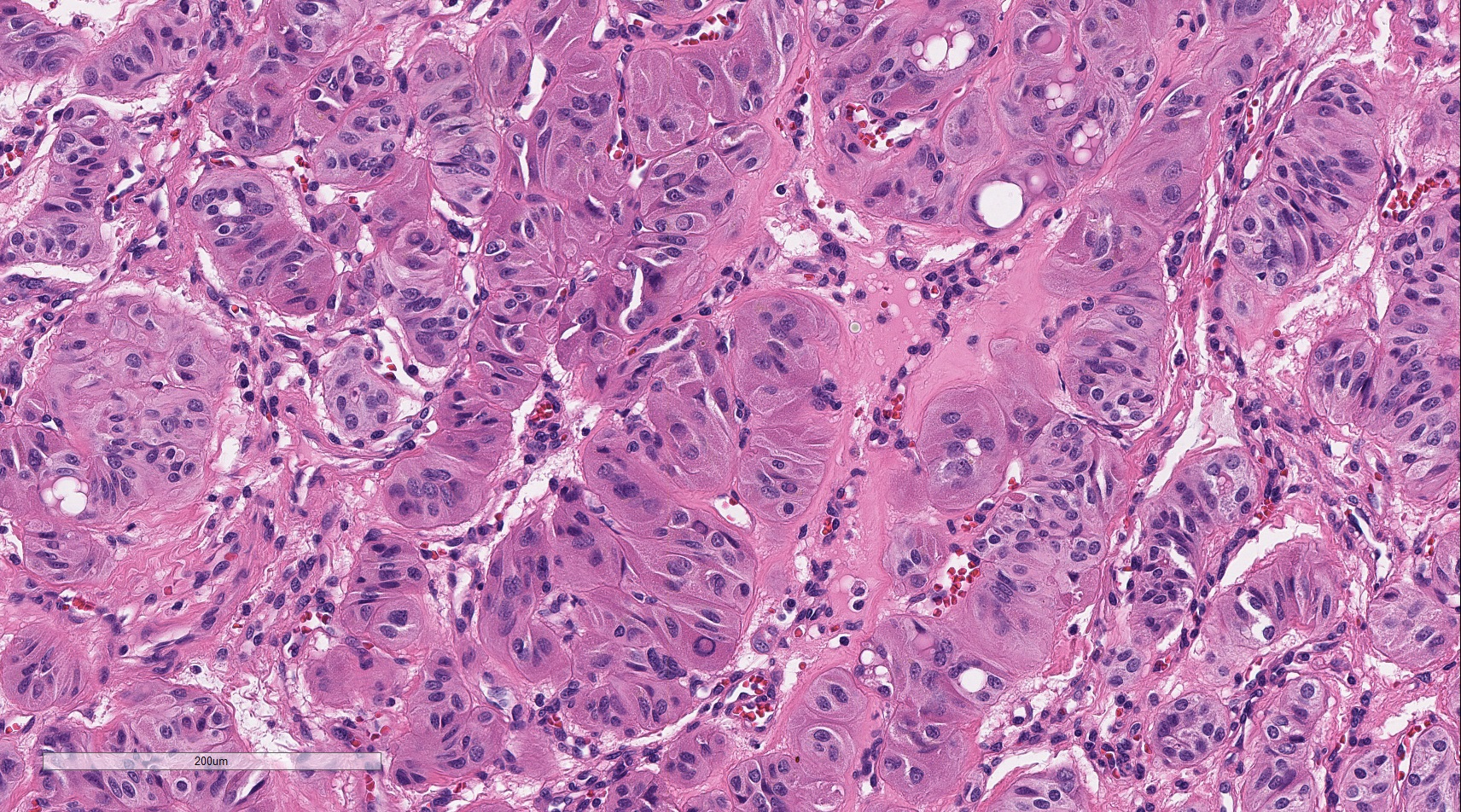
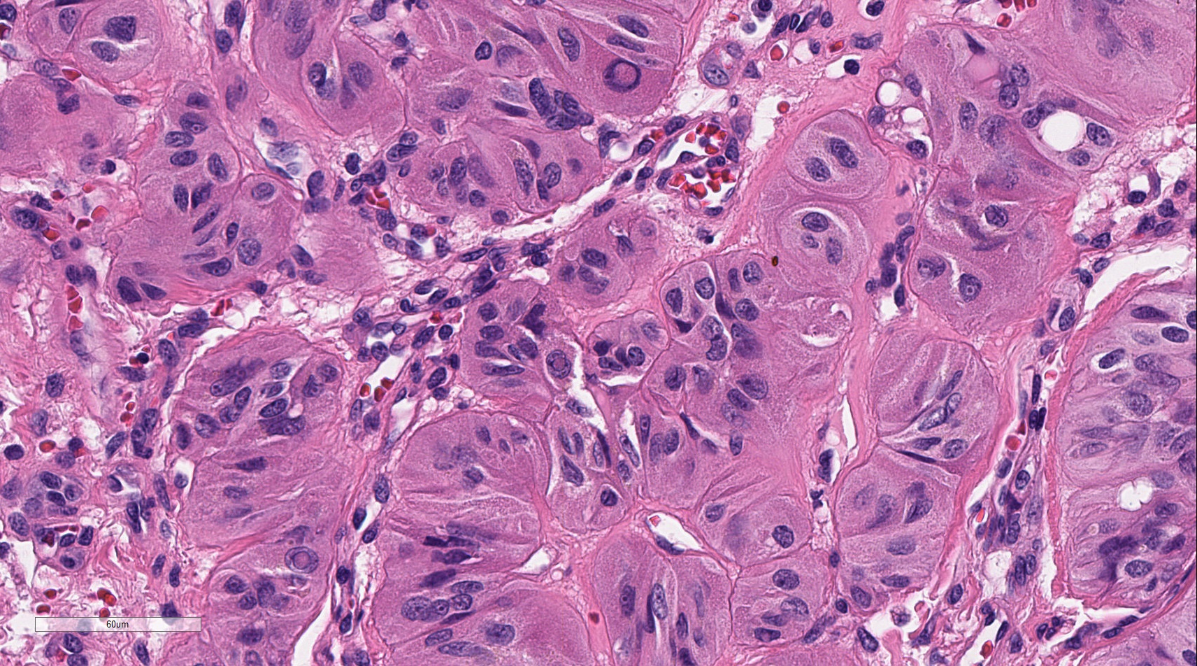
The patient is a 43 year old woman and navy veteran who was brought to the attention of the Mather Veterans Affairs otolaryngology service after a CT scan conducted as part of a routine trauma survey following a motor vehicle accident revealed a nodular thyroid. Subsequent ultrasound of the right lobe (4.3 x 2.3 x 1.9 cm) demonstrated a right inferior-medial solid slightly hypoechoic nodule (1.5 x 1.2 x 0.8 cm), an inferior lobe solid heterogeneously hypoechoic nodule (1.1 x 1.0 x 1.0 cm), and a lateral right mid lobe solid hypoechoic nodule with some vascularization (0.9 x 0.9 x 0.9 cm). FNA sampling of the 1.1 cm nodule was conclusive for papillary thyroid carcinoma while sampling of the 1.5 cm nodule revealed an atypia of unknown significance. Permanent sections of the 1.5 cm nodule revealed a well circumscribed neoplastic process with cells arranged in a predominantly trabecular pattern with prominent nuclear grooving,scattered nuclear pseudoinclusions, and abundant amounts of hyalinized cytoplasm in a background of chronic lymphocytic thyroiditis.
Microscopic Photographs:
 |
 |
|
| 01x | 04x | |
 |
 |
|
| 10x | 20x | |
 |
||
| 40x |

 Meet our Residency Program Director
