
Residency Program - Case of the Month
May 2016 - Dr. Trevor Starnes
History:
The patient is a 55-year-old woman with a history of alcoholic cirrhosis, currently being treated for breast cancer, who noticed a slowly-growing left cheek mass one year ago. She denies having pain or any other symptoms associated with this mass until the past month as the mass began to ulcerate through the skin. The surgeons treated her using a left superficial parotidectomy with wide local excision of skin and level 2 selective neck dissection.
Gross Findings:
The skin contains a centrally located, puckered defect (0.4 cm in greatest dimension).
The specimen is serially sectioned to reveal a slightly firm, pink-white, fibrous appearing area (1.3 x 1.3 x 1.0 cm) located immediately subjacent to the puckered defect on the skin surface.
Microscopic Findings:
This is a cystic mass with predominance of well differentiated mucinous cells mixed with a component of squamous-like intermediate cells. Mucinous cells are large with basally displaced nuclei.
Click to enlarge
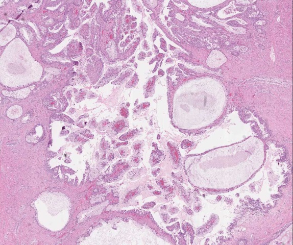
Figure 12x view of mass |
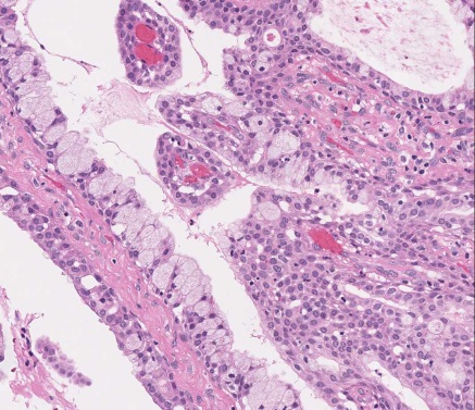
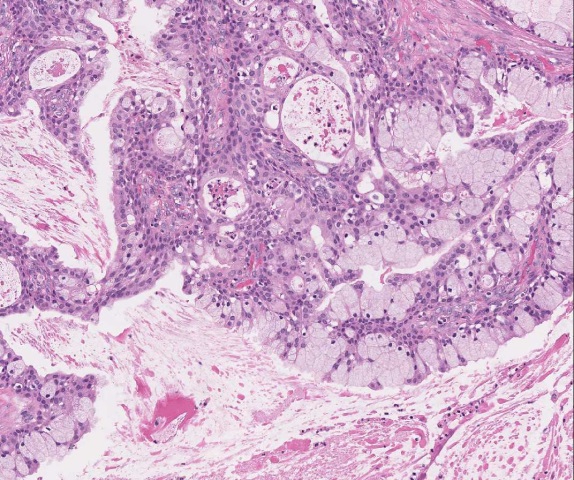
Figure 210x view of mass |
Figure 320x view of mass |
||
|
|
|
 |
What is the diagnosis?
Choose one answer and submit.
 Meet our Residency Program Director