Residency Program - Case of the Month
July 2014 - Presented by Jessica Rogers, M.D. & Pritesh Chaudhari, M.D.
Clinical History:
The patient is a 35-year-old female with a medical history significant for hypothyroidism, depression, and asthma. She presented to her primary care physician in January 2014 with four days of fever and cough after having returned from a trip to Egypt and Israel for which she was placed on antibiotics. Due to the persistence of her cough, in early March 2014 a chest x-ray was ordered, which showed a rim calcified mass in the left upper quadrant of the abdomen. A subsequent abdominal CT showed an 8.7 cm cystic splenic mass with peripheral calcification and a 5.1 cm splenic soft tissue density. An MRI on April 2, 2014 showed several splenic lesions with varying degrees of solid and cystic components measuring up to 8.7 cm in diameter. Due to the patient’s travel history to Nicaragua, Laos, Cambodia, and Thailand, the differential diagnosis included a post-infections process (including Echinococcus), Lymphangioma, Hemangioma, and malignancies. Echinococcal antibody tests were performed on March 11, 2014 and May 30, 2014, which were negative. The patient was seen by infectious disease specialist in San Francisco and subsequently started Albendazole. She was thought to be improving but continued to have a cough and left upper quadrant pain in addition to night sweats, weight loss, and abdominal fullness. Over the next couple months, the patient developed a worsening leukocytosis, microcytic anemia, and thrombocytosis. Labs obtained June 30, 2014 again showed a WBC of 22, Hematocrit of 21, and thrombocytosis to a platelet count of 667. Thus, she was admitted for treatment and ultimately underwent a splenectomy.
Gross Description:
Received fresh for lymphoma workup and subsequently placed in formalin designated "spleen," is a 980 gram, 16 x 15 x 9 cm spleen with a large 15.0 x 9.0 x 8.0 cm cystic mass. There are several 1.2 to 2.0 cm lobulated small mass protruding from the large cytic mass. The cystic cavity is surrounded by thick wall with peripheral calcifications. The cystic cavity contains cloudy, yellow fluid (approximately 40 mls) with suspended soft necrotic debris. The solid area of the mass is well defined, tan-pink and firm. The remaining splenic parenchyma is pink to red, firm, and the splenic capsule appears intact. No hilar lymph nodes are identified.
Gross Images:
 |
 |
|
 |
 |
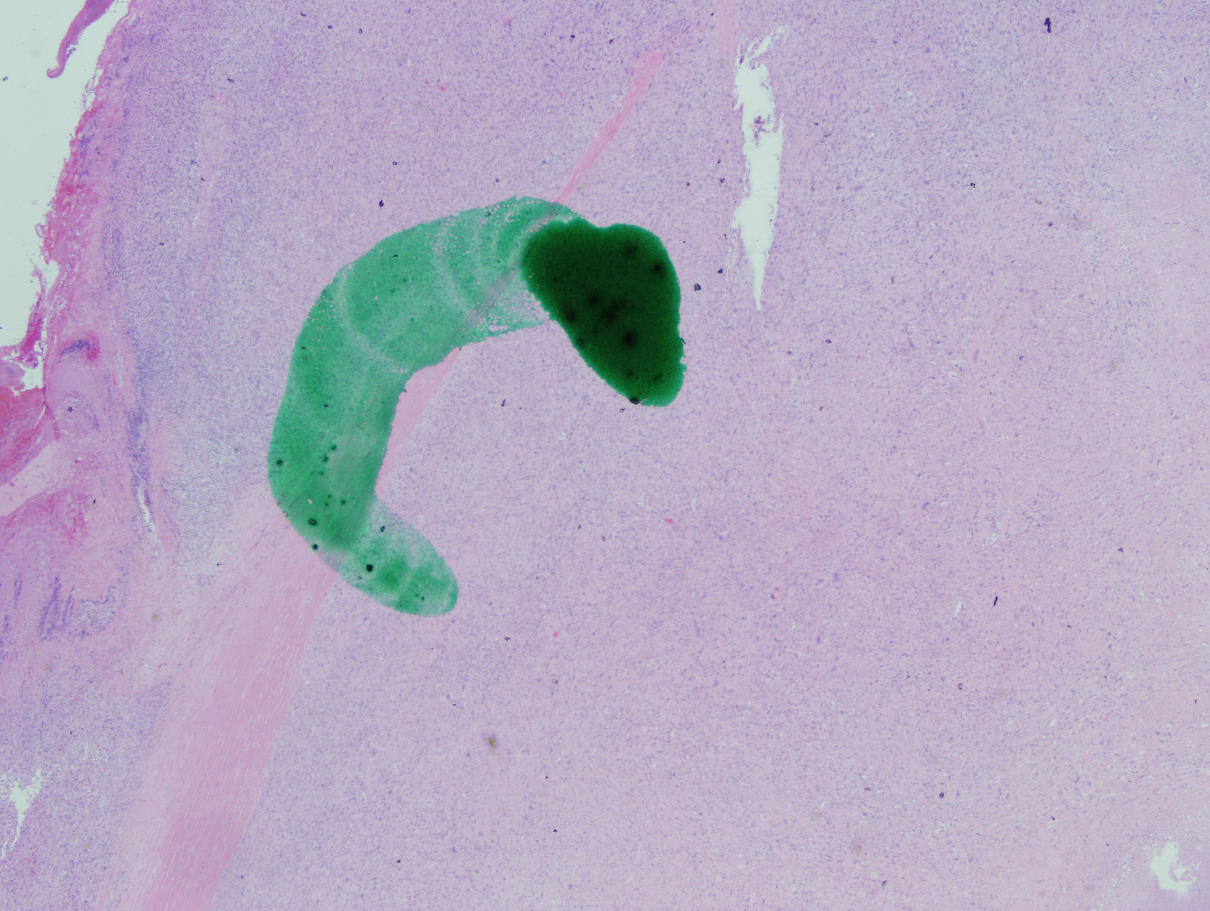
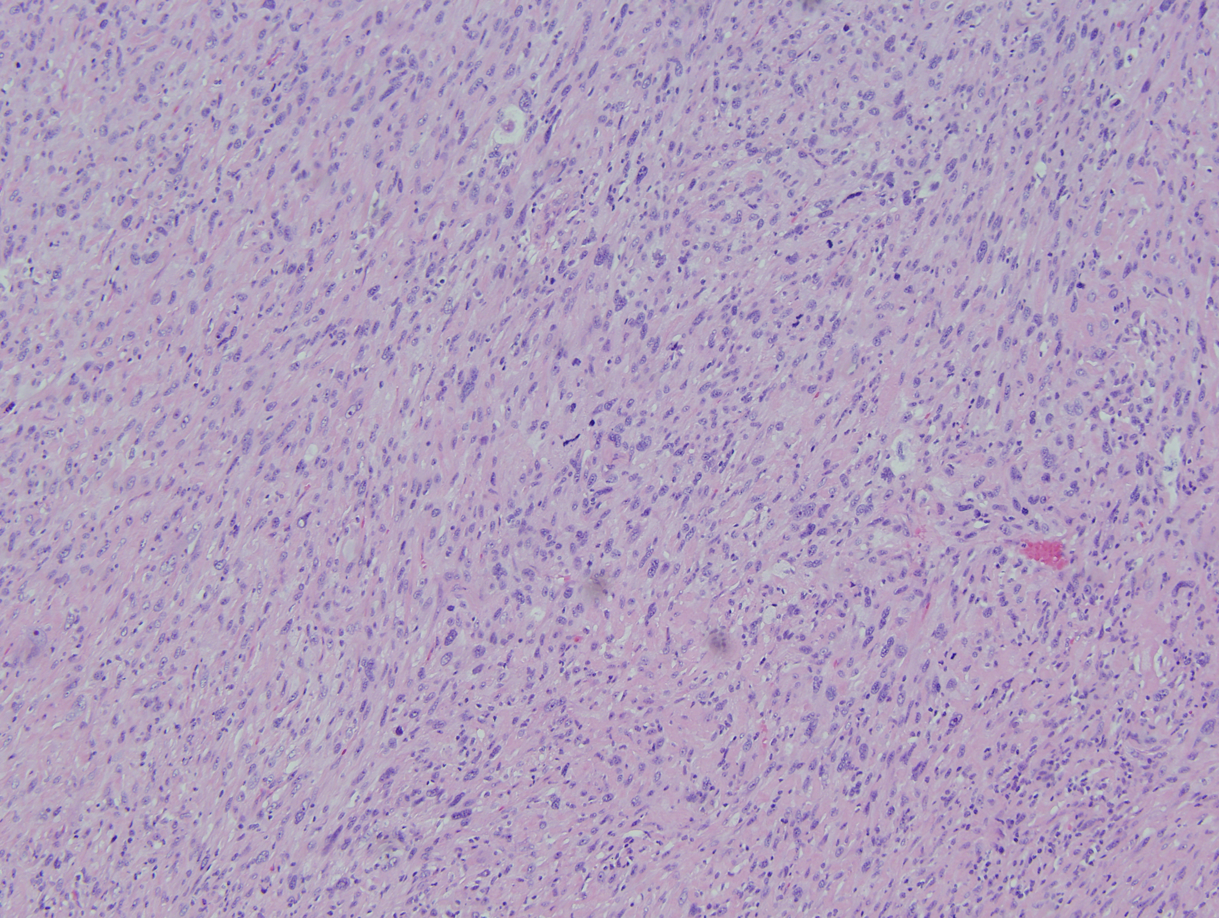
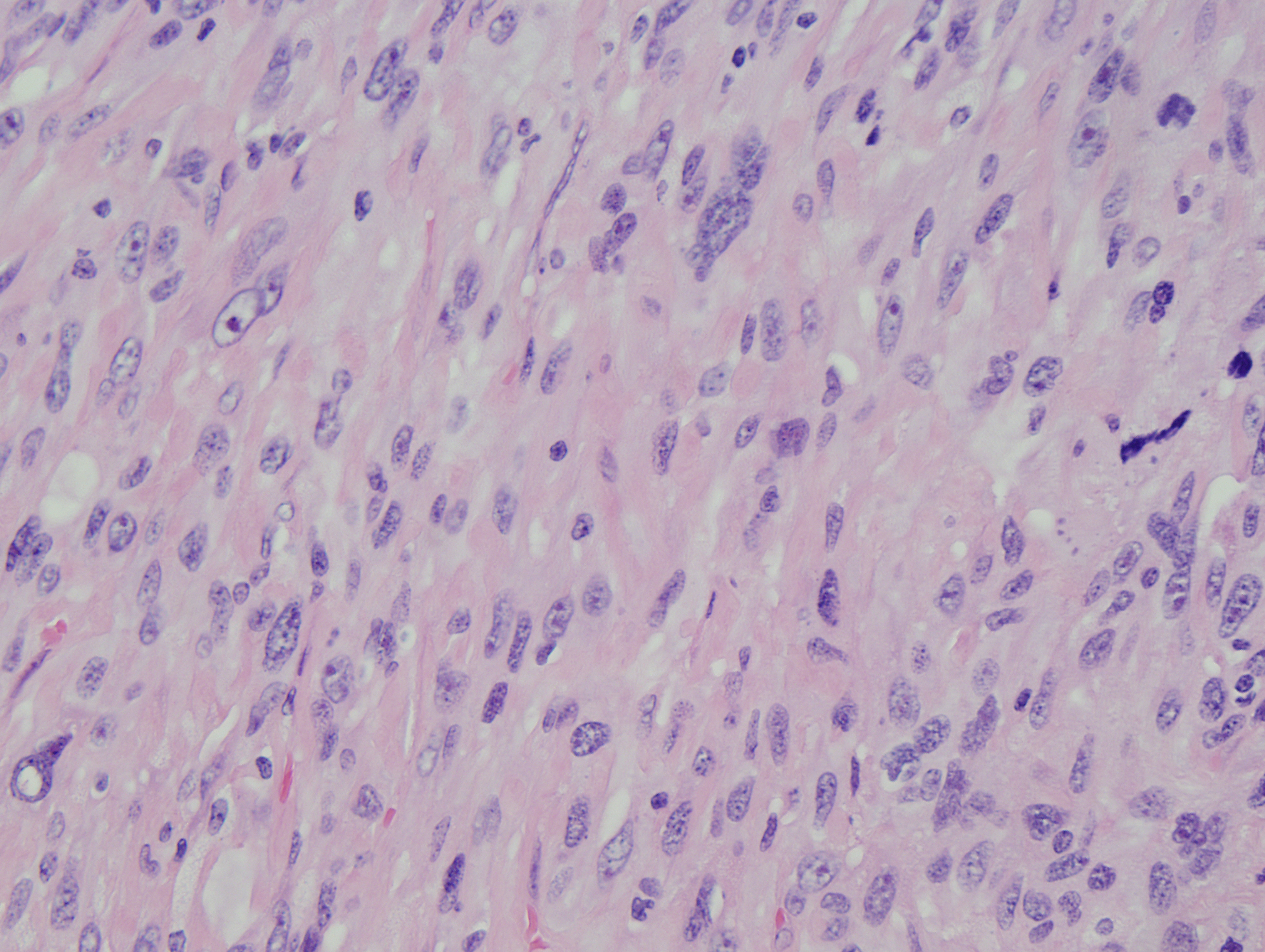
Microscopic Images:
 |
 |
 |
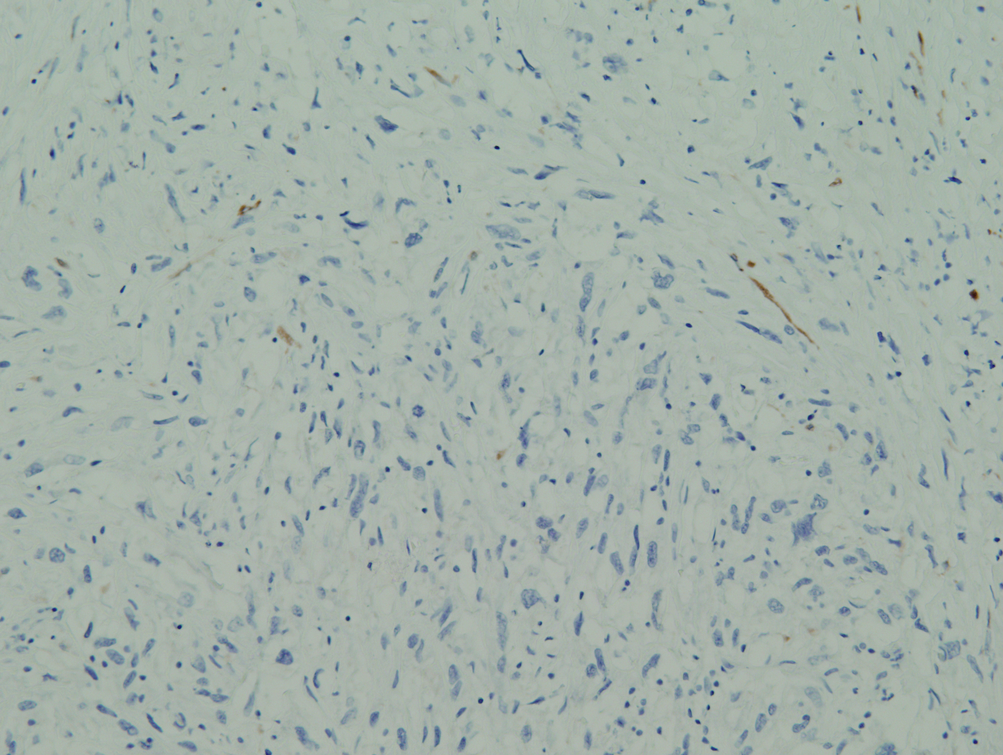
Immunohistochemistry:
 |
 |
 |
||
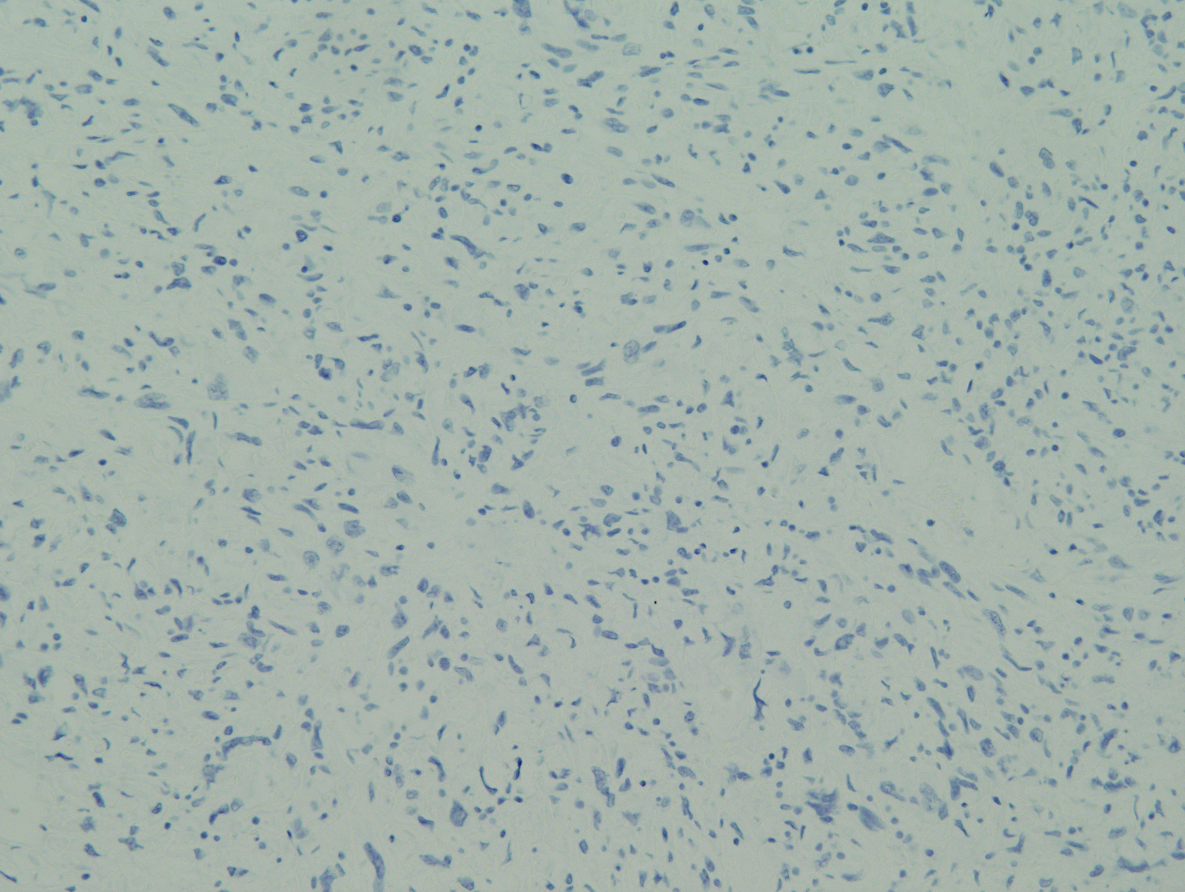
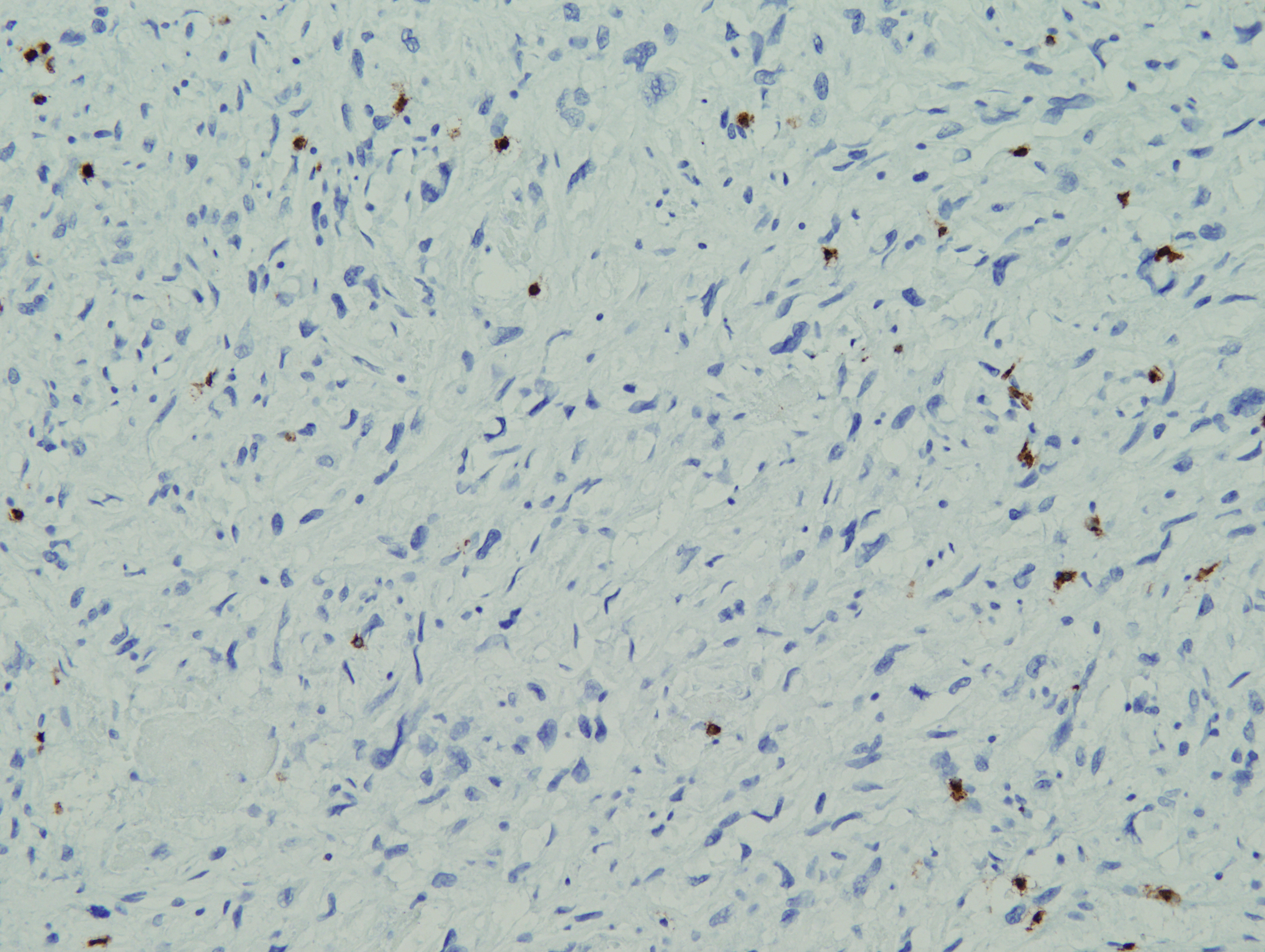
| AE1/AE3 | HMB45 | CD3 | ||
 |
 |
 |
||
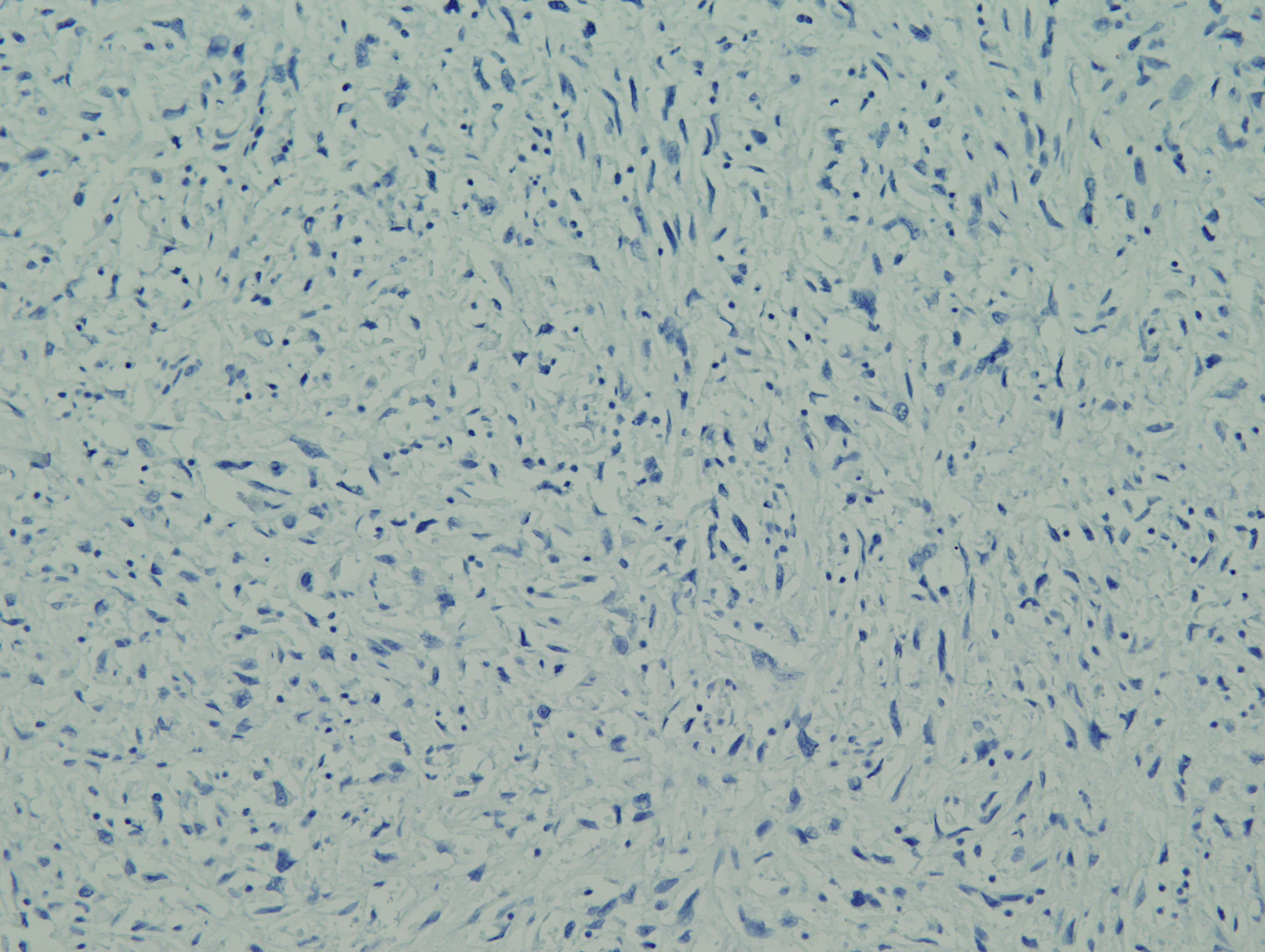
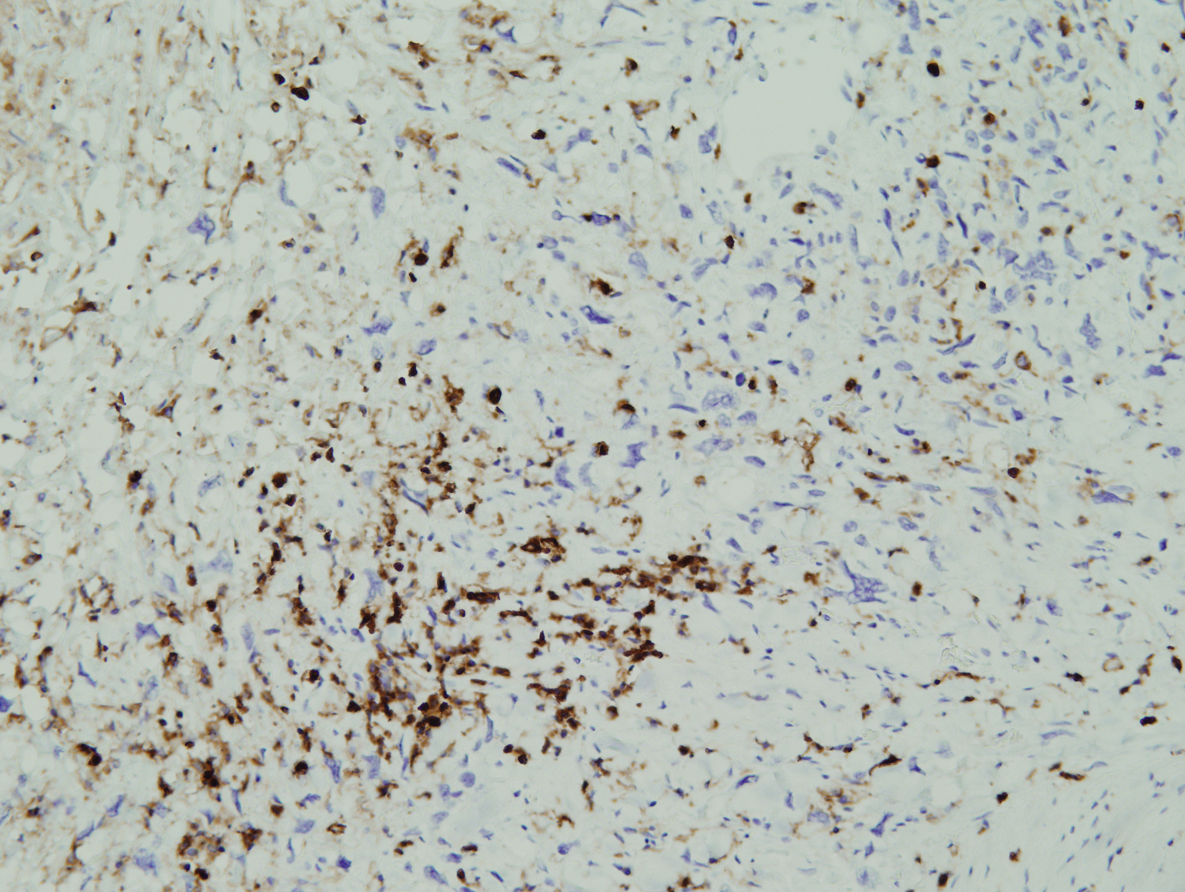
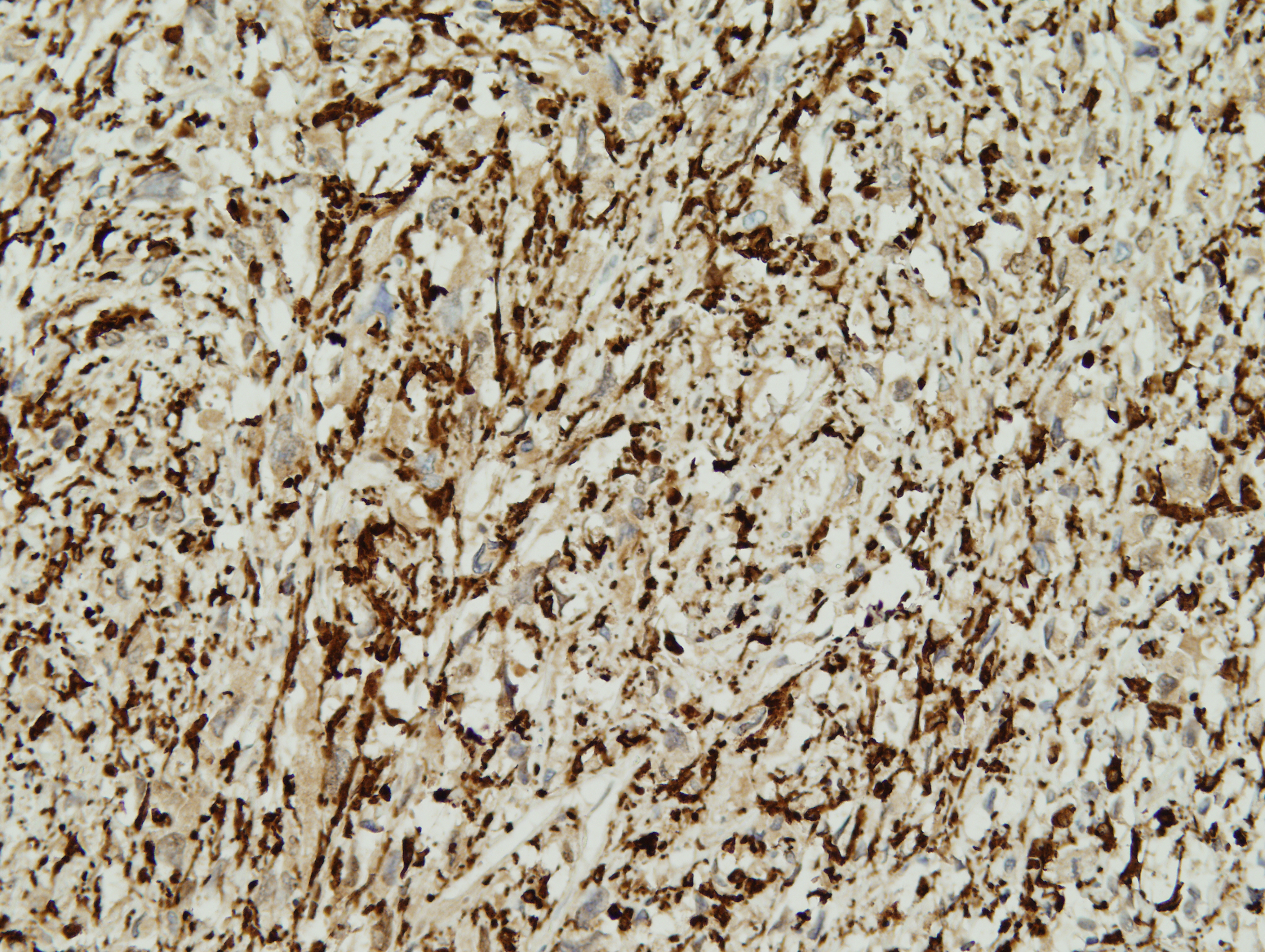
| PAX5 | CD45 | CD163 | ||
 |
||||
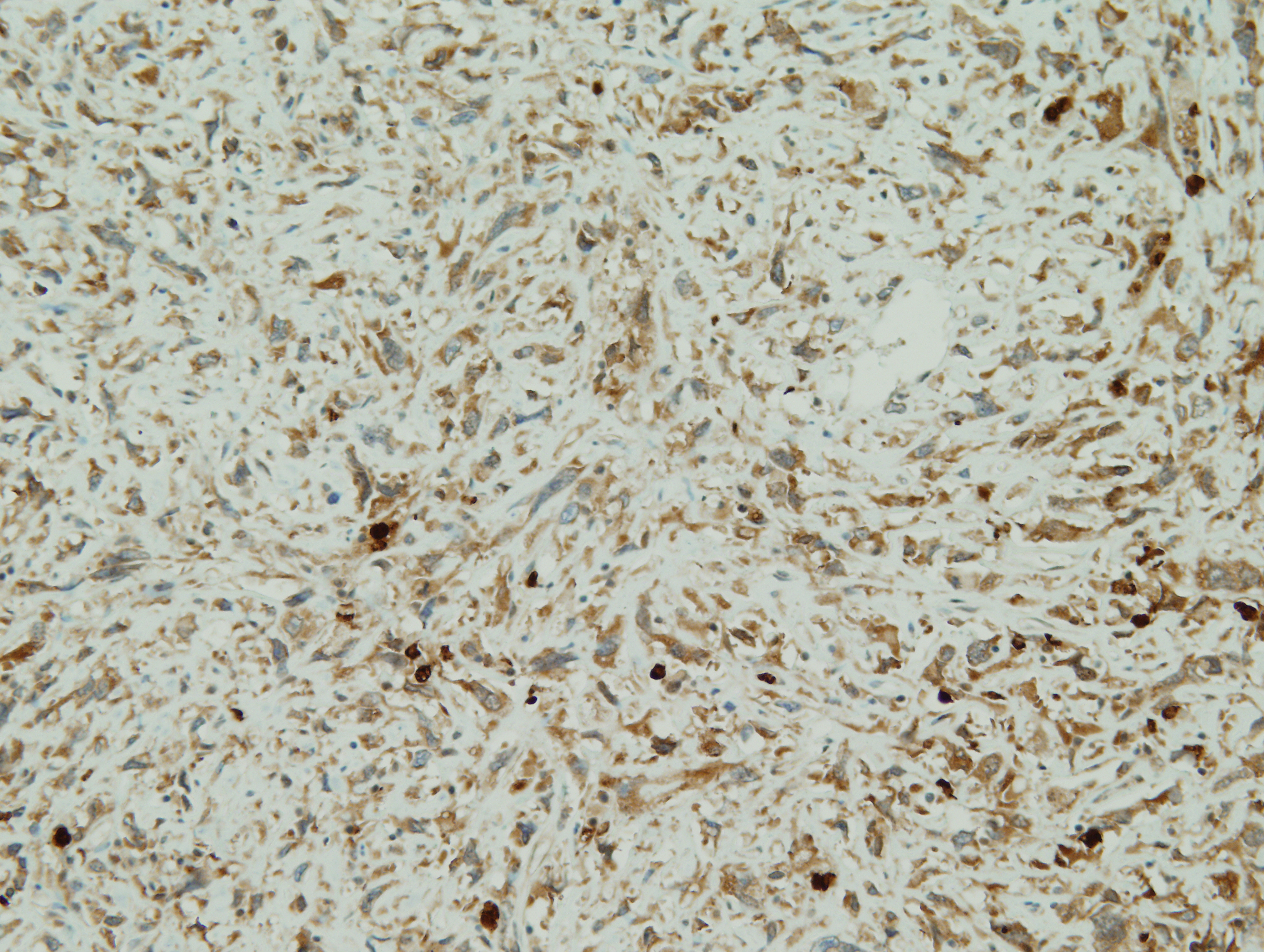
| Lysozyme |
Answer

 Meet our Residency Program Director
