Cause of common type of heart failure may be different for women and men
By Lisa Howard
Mouse study identifies sex differences at the cellular level for heart failure with preserved ejection fraction (HFpEF)
(SACRAMENTO)
A new study from the UC Davis School of Medicine found striking differences at the cellular level between male and female mice with heart failure with preserved ejection fraction (HFpEF).
The findings could determine how HFpEF is treated in women compared to men.
With HFpEF, the heart muscle contracts normally but the heart is unable to fully relax and refill properly between beats. This condition is known as diastolic dysfunction. It can occur if the heart is too stiff or if the contraction process doesn’t shut off quickly enough between beats.
The study showed that the diastolic dysfunction in female mice resulted from altered heart filament proteins. In male mice, it resulted from the slow removal of calcium from heart cells between heartbeats, causing a slight contraction to remain between beats.
“This study demonstrates the importance of conducting research on both male and female populations,” said Donald M. Bers, a senior author of the study. Bers is the chair of the Department of Pharmacology and the Joseph Silva Endowed Chair for Cardiovascular Research at the UC Davis School of Medicine. “If these same molecular male-female distinctions occur in obese diabetic patients with HFpEF, it may mean that the best therapeutic strategies for HFpEF in women may differ from those for men.”
If these same molecular male-female distinctions occur in obese diabetic patients with HFpEF, it may mean that the best therapeutic strategies for HFpEF in women may differ from those for men.” —Donald M. Bers, chair of the Department of Pharmacology and the Joseph Silva Endowed Chair for Cardiovascular Research
“Two hit” mouse model to study HFpEF
Obesity and diabetes are common in people with HFpEF. To study the disease, the researchers created a unique “two-hit” mouse model combining two factors.
For the first factor, the researchers used mice genetically lacking a leptin receptor. Leptin is a hormone that promotes satiety. Without it, appetite remains high and the animals become obese and diabetic. For the second factor, mice were exposed to an aldosterone infusion. Aldosterone is a hormone made by the adrenal gland. High levels of aldosterone cause fluid retention.
This animal model of heart failure and diabetes develops HFpEF, allowing researchers to analyze the cellular and molecular mechanisms of muscle contraction and relaxation in male and female mice.
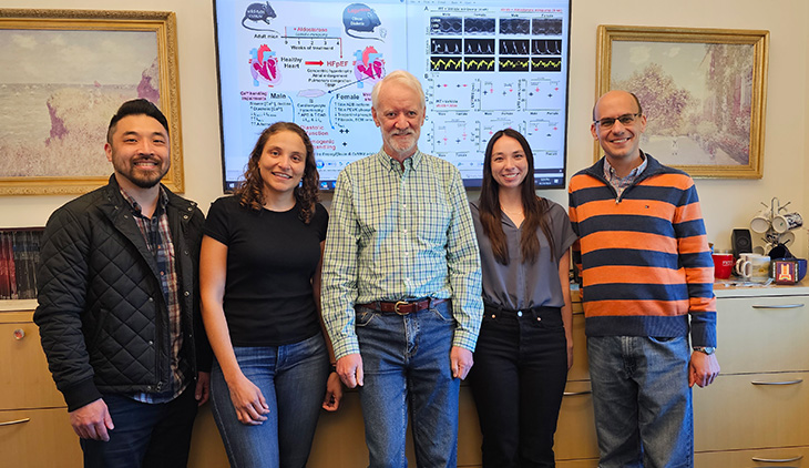
The research team (left to right): Christopher Y. Ko, Juliana Mira Hernandez, Donald M. Bers, Erin Y. Shen and Bence Hegyi in in front of their key findings on the screen.
Dysregulation of calcium, titin
Calcium is critical in the activation of contraction and relaxation of heart muscle cells as well as the heart’s electrical activity. Calcium entering the heart cell at each beat causes the muscle to contract. It also helps drive the electric signal that synchronizes the contraction of the millions of heart muscle cells required for the heart to function as an efficient pump. Calcium is removed from the cell at each beat. This allows the heart to relax between beats and fill for the next beat.
In the male mice with HFpEF, the calcium removal from the heart muscle cells was slowed, preventing complete relaxation between beats. The male HFpEF mice also exhibited more abnormal heart rhythms, known as arrhythmias.
In contrast, the females with HFpEF exhibited normal calcium movements into and out of the heart cells. Instead, the researchers observed an increase in a shorter and stiffer form of titin (N2B). Titin is a protein in the heart that acts like a supportive spring. Researchers also observed phosphorylation (a molecular reaction) of titin and another heart filament protein, troponin I. Both the titin and troponin changes made the female heart cells functionally stiffer — making the heart harder to fill — even though calcium removal was normal.
“This study reveals different drug targets in males and females and will be a stepping-stone for future trials with sex-specific targeted drugs in HFpEF,” said Bence Hegyi, an associate project scientist in the Bers Lab and co-senior author of the study. “Potentially, women with this form of HFpEF could benefit from drugs that reduce cardiac stiffness. On the other hand, men with this form of HFpEF might benefit more from drugs that enhance calcium removal.”
Limitations of the research
The researchers noted several limitations of the study. Although the mice in this study may be representative of the substantial number of HFpEF patients who have diabetes and are quite obese, many HFpEF patients may not be represented by this model. Multiple animal models will be needed to understand different subpopulations with HFpEF. Additional preclinical and clinical studies are needed to fully realize the potential benefits of this work.
Additional authors include Erin Shen, Christopher Ko, Emily Spencer, Daria Smoliarchuk and Julie Bossuyt from the UC Davis School of Medicine; Juliana Mira Hernandez from the UC Davis School of Medicine and the University of Antioquia, Medellin, Colombia; and Zaynab Hourani and Henk Granzier from the University of Arizona, Tucson.
Funding: The project was funded by the National Institutes of Health grants (P01-HL141084, R01-HL142282, and R35-HL144998); the American Heart Association Career Development Award 23CDA1051603; the Stanford Diabetes Research Center grant P30DK116074, and the Minciencias – Fulbright Colombia Scholarship.