UC Davis develops engineered bone marrow to study early osteosarcoma progression
Researchers test interactions of macrophages with bone cancer cells in a new 3D bone marrow model
(SACRAMENTO)
UC Davis pediatric orthopedic surgeons, biomedical engineers, and immunologists have created an artificial bone marrow model that may help them better understand osteosarcoma, the most common type of bone cancer in children and adolescents.
The model will allow them to study the effects of different types of special immune cells – known as macrophages – on the progression and spread of the disease.
Osteosarcoma, which typically originates in the bone marrow, mostly develops in bones around the knee and upper arm but can spread to other parts of the body. Its treatment has barely changed in the last four decades. The current 5-year survival rate of people with a spreading osteosarcoma is less than 25%.
“There is a great need for reliable predictive indicators and treatments for this disease,” said Katherine Griffin, a doctoral candidate in immunology and veterinary medicine and researcher in the Leach Laboratory at the UC Davis Department of Orthopaedic Surgery. “And for that, we need to find new models of osteosarcoma to boost our understanding of its progression and the way immune cells like macrophages interact with the cancerous cells.”
Engineered bone marrow as host for osteosarcoma cells
Scientists rely on animal models that can mimic aspects of a disease found in humans. These models help them study disease prevention, diagnosis, development and treatment.
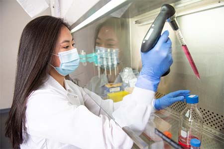
Katherine Griffin at Leach Laboratory (Photo by Don Preisler)
Osteosarcoma grows in a complex bone marrow environment. Current models do not account for key components of this environment, including the 3D nature of sarcomas and the physiological oxygen tension of bone marrow. Both components could impact the disease’s behavior.
The UC Davis team constructed engineered bone marrow (eBM) that mirrors the heterogeneous cell population found in native bone marrow and provides a new 3D platform to study tumor progression.
“We place elements that naturally make bones – such as a supportive bony matrix, special proteins that direct cells to become bone cells, and collagen– in small molds and implant them into the back of a mouse model. We let the implant grow for eight weeks as the mouse’s own cells populate the mold. We then collect those constructs and maintain them in culture to perform osteosarcoma research studies,” Griffin explained.
Osteosarcoma is also an extremely heterogeneous cancer, both from patient to patient and within an individual patient. Thus, using a single cell line has major limitations.
While the idea of studying macrophages and how they affect tumors is not a novel concept, we are innovating in this space by applying a tissue-engineered platform toward a cancer immunology question.”—Kent Leach
Griffin noted that most osteosarcoma studies are done with only one cell type in standard culture in ambient air (21% oxygen). While the physiological oxygen-tension of bone marrow is 5%, most, if not all, experiments take place at elevated 21% oxygen levels. Their new studies will examine how low oxygen (hypoxia) affects tumor progress in the bone marrow.
“We are trying to establish a new model and show that it's more accurate than the typical monolayer culture for cancer cells,” Griffin added.
Macrophages and osteosarcoma
Previous studies have linked macrophages to osteosarcoma progression.
Macrophages, a diverse group of white blood cells, are formed in response to an infection or accumulating damaged or dead cells. Their role is to detect and destroy defective cells and harmful organisms like bacteria and viruses. They can cause inflammation by releasing molecules known as cytokines.
The team will introduce osteosarcoma cells both with and without adding macrophages to their engineered bone marrow samples. Then, they will use special dyes to stain the sarcoma and macrophage cells and track the movement of these cells over time.
As the macrophages play a key role in shaping the tumor microenvironment, their infiltration may correlate with the disease development. Yet, the influence of macrophage phenotypes on primary and metastatic osteosarcoma behavior and disease progression is still unknown. The team hypothesizes that the polarization of one phenotype known as M2 macrophage will cause osteosarcoma growth within the bone marrow model.
“While the idea of studying macrophages and how they affect tumors is not a novel concept, we are innovating in this space by applying a tissue-engineered platform toward a cancer immunology question. There are few groups attempting to model cancer in the dish with the remarkable complexity observed in the body,” said Kent Leach, professor of orthopedic surgery and biomedical engineering at UC Davis and an expert in biomaterials. “The intersection between cancer biology, immunology, tissue engineering, and patient care evident in this project is a rare combination!”
This research is a collaboration among UC Davis researchers, including Lor Randall, an internationally recognized sarcoma surgeon and scientist, and Steven Thorpe, assistant professor of pediatric orthopedic surgery.