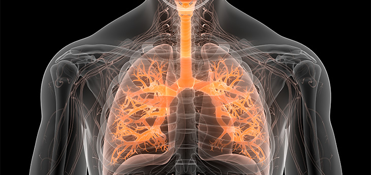
Lung cancer breakthroughs: New imaging technologies may change course for top cancer killer
By Stephanie Winn
High tech 3D imaging spots early lung cancer while another tool tracks lung cancer treatment
(SACRAMENTO)
UC Davis Health is first in the region to deploy two new technologies that bring hope to people with lung cancer. Both provide advanced imaging. One helps catch lung cancer earlier and the other tracks the progress of lung cancer treatment.
Technology No. 1: New 3D CT imaging system helps diagnose lung cancer earlier
A new state-of-the-art mobile 3D CT imaging system is catching lung cancer earlier by pinpointing potentially cancerous growths for more precise lung cancer biopsies.
The lung moves as people breathe, which makes it challenging to navigate during biopsies. UC Davis pulmonary doctors looked for a way to improve the odds of finding lung cancer at an earlier stage. They decided to pair the Cios Spin, made by Siemens Healthineers, with a breakthrough robotic-assisted bronchoscopy system called the Ion, made by Intuitive.
It’s similar to driving your car and using the GPS system to update your destination. In this case, we are using Cios Spin to update the exact location of the lung nodule biopsy.”—Chinh Phan, director, Interventional Pulmonology Program
Robotic-assisted bronchoscopy allows doctors to precisely examine air passages with a controller that operates a small camera at the end of a flexible tube. The Cios Spin aids this advanced tool by using cone beam computed tomography to create real-time 3D images. The images automatically update the best path to lung nodules or lesions.
“The synergy of these two systems improves the precision and accuracy of lung nodule biopsies,” said Chinh Phan, director of the UC Davis Health Interventional Pulmonary Program. “This enables safer and earlier diagnosis of lung cancer.”
How it works
Phan said he first uses the Ion robotic-assisted bronchoscopy to guide an ultra-thin catheter to reach the outer areas of the lung. Cios Spin then provides real-time cone beam CT images to confirm the exact location of the lung nodule.
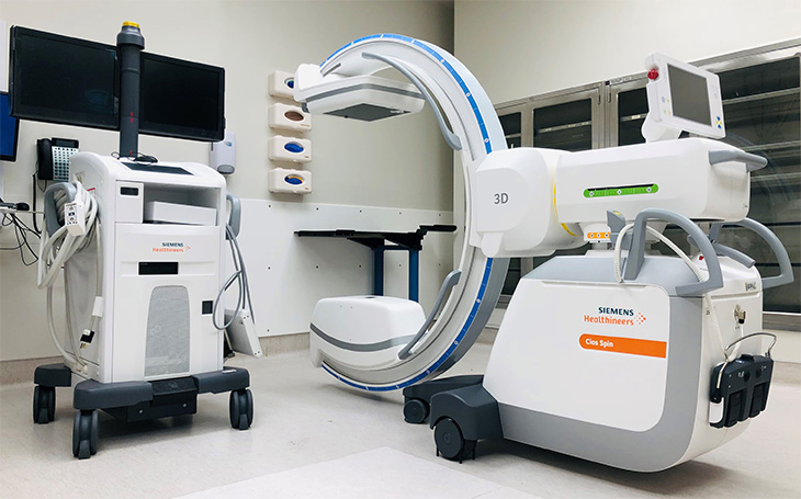
Mobile 3D CT imaging system Cios Spin
The Cios Spin communicates with Ion instantly. The CT data is transferred right away to Ion during the procedure. This enables the doctor to immediately re-target the lung nodule for a more precise biopsy.
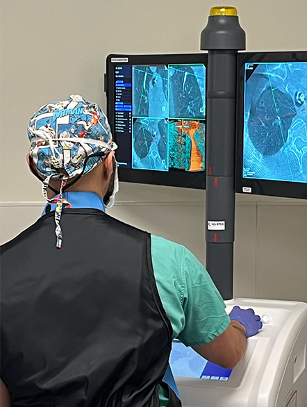
Chinh Phan using the CisoSpin during a robotic-assisted bronchoscopy
Images in real time
This real-time visualization means shorter procedures, improved accuracy and safer biopsies.
“It’s similar to driving your car and using the GPS system to update your destination. In this case, we are using Cios Spin to update the exact location of the lung nodule biopsy,” Phan said.
Results show lung cancer is being caught earlier when it is more treatable
After a year of using the Cios Spin and Ion together, Phan said he and his team have seen a major shift. Lung cancer is being diagnosed much earlier when it is more treatable. In fact, for the first time, he said that UC Davis Comprehensive Cancer Center is seeing more lung cancers diagnosed at stage 1 as compared to late stage.
“The results using these combined technologies that pairs advanced CT imaging with robotic bronchoscopy is transforming lung cancer care for the patients in our community,” Phan said.
Technology No. 2: Advanced imaging may hold key to tracking lung cancer treatment
Lung cancer can advance quickly. Once a person starts treatment, it is vital to know if that treatment is working or if the patient should be switched to a different treatment. New advanced imaging technology at UC Davis Health may give doctors a way to quickly track how well their patients are doing with a certain treatment.
Scientists at UC Davis Comprehensive Cancer and the UC Davis Department of Radiology have discovered a novel way to image the unique blood supply of the lungs. Their findings suggest it could help determine if lung cancer treatment is working.
Pioneering EXPLORER gives scientists a peek inside the workings of the lungs
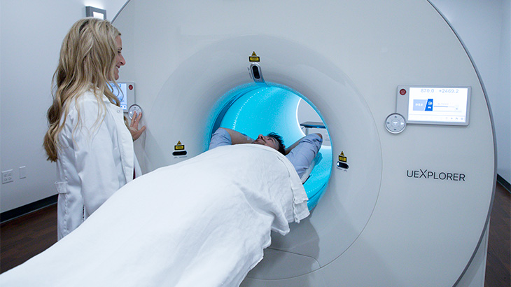
The groundbreaking research uses EXPLORER, an innovative PET scanner invented at UC Davis and housed at the UC Davis Health Molecular Imaging Center in Sacramento. EXPLORER provides images of the body in minutes with higher quality and less radiation than traditional positron emission tomography (PET) scanners.
Our study found that the dual-blood supply effect is bigger in lung tumors than in normal lung tissue, which is why it can be used to spot lung cancer and has the potential to help us understand how it spreads.”—Guobao Wang, professor, Department of Radiology
Findings from the new lung cancer study were published in the Journal of Nuclear Medicine. They how for the first time how fast dynamic imaging using an advanced PET scanner, such as EXPLORER, can “see” into organs that have a dual blood supply. The lungs receive deoxygenated blood from the pulmonary arteries and oxygenated blood from the bronchial arteries.
Patient entering advanced PET scanner EXPLORER
Viewing the dual blood supply
Cancer frequently spreads to the lungs from other parts of the body because of this extensive blood supply.
“Our study found that the dual-blood supply effect is bigger in lung tumors than in normal lung tissue. This is why it can be used to spot lung cancer and has the potential to help us understand how it spreads,” said Guobao Wang, a professor with the Department of Radiology, who is leading the study.
Insights from the study
Wang added that lung cancers often don’t do well with treatments such as immunotherapy. Analyzing lung tumors with a powerful PET imaging tool may lead to better therapy.
“In this new work, we are combining high frame-rate movies from EXPLORER with advanced math to develop a new PET imaging method. This makes it now possible to see and understand the dual-blood supply in normal lung tissue and lung tumors,” added Yiran Wang, a doctoral candidate in the Wang Lab and the first author of the research paper. He was jointly supervised by Simon Cherry, one of the co-inventors of the EXPLORER scanner.
Why it matters
Lung cancer is the leading cause of cancer death in the United States. An estimated 125,000 Americans will lose their lives to the devastating disease this year.
UC Davis Comprehensive Cancer Center is the only National Cancer Institute-designated center serving the Central Valley and inland Northern California, a region of more than 6 million people. Its specialists provide compassionate, comprehensive care for more than 100,000 adults and children every year and access to more than 200 active clinical trials at any given time. Its innovative research program engages more than 240 scientists at UC Davis who work collaboratively to advance discovery of new tools to diagnose and treat cancer. Patients have access to leading-edge care, including immunotherapy and other targeted treatments. Its Office of Community Outreach and Engagement addresses disparities in cancer outcomes across diverse populations, and the cancer center provides comprehensive education and workforce development programs for the next generation of clinicians and scientists. For more information, visit cancer.ucdavis.edu.