Mixed reality goggles, 3D printing aid in surgical planning to separate craniopagus twins
Conjoined twins Abigail and Micaela Bachinskiy were born connected at the skull and brain. It’s a rare condition called craniopagus twins which occurs once in every 2.5 million births.

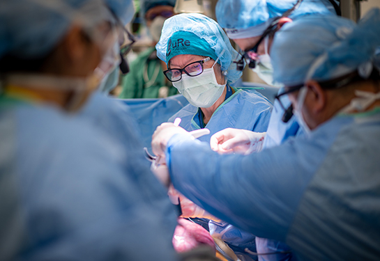
At nine months old, Abigail and Micaela were successfully separated in a marathon surgery at UC Davis Children’s Hospital on Oct. 24 and 25, 2020. It was the culmination of months of planning and intense preparation that would be – for most of the surgical team – the most complicated case of their careers.
“No two conjoined twins born are the same. Each case is different. Their anatomy is different. There are no textbook models of how you separate the twins,” said Michael Edwards, lead pediatric neurosurgeon on the case.
But technology was on their side. From mixed reality imaging goggles that map out the brain and blood vessels to three-dimensional 3D printed models of their skulls, the team had access to equipment and technology that did not exist 10 years ago to help them plan and practice this surgery with precision.
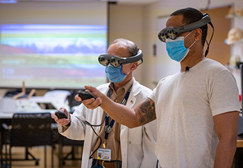
Mixed reality goggles help visualize operation plan
The surgical team spent months carefully tracking the twins’ growth through MRI and CT scans, which revealed that the twins shared some bone, brain, blood vessels and soft tissue.
Mixed reality goggles used this imaging data to create a three-dimensional view inside the twins’ skulls. The surgery team had a vantage point into the babies’ unique anatomy from every possible angle.
“You can look from the top, the side, the bottom, you can rotate the 3D model. You can walk into it and look backward to see where you are,” Edwards said.
A view inside the goggles showed a complex network of blood vessels which the team would need to detangle and separate during the separation surgery.
“By working in three dimensions, we have a better idea of what things are actually going to look like when we are in surgery. It adds a significant margin of safety,” Edwards said. “With these new techniques, we have the ability to view the anatomy from any perspective, wipe away the bone, look at the dura. Wipe away the dura and look at the brain and vasculature.”
The team could plan the operation in three dimensions on this virtual system and identify potential pitfalls without risk to the babies. They could also rehearse working in complicated areas, so they knew what to expect in the operating room.
3D printing of their skulls

Michael Edwards and Granger Wong examine the 3D model of the twins’ skulls.
Harnessing the power of three-dimensional printing, the team was able to use three-dimensional models of the twins’ skulls and blood vessels to assist in their planning.
“The alternative is a two-dimensional picture. That automatically requires the surgeon and the team to have the ability to conceptualize what this would be like in three dimensions. It’s not so easy to understand,” Edwards said. “We’re working with a 3D baby in a 3D world. Two-dimensional images don’t provide us with all the information that we need or can use.”
Private vendor KLS Martin made custom 3D printed models of the twins’ skulls that were primarily used by UC Davis Children’s Hospital team, based on CT and MRI scan information. UC Davis Children’s Hospital also received models from the 3D PrintViz Lab, a state-of-the-art facility on the UC Davis Sacramento campus.
“You can see how the twins are connected. They are asymmetrical. They are not joined back to back. It’s more of a side to back configuration,” said Granger Wong, chief of the UC Davis Division of Plastic and Reconstructive Surgery and lead plastic surgeon on the twins’ case. “Luckily we exist in a time with this technology.”
Based on these models, Wong knew the exact amount of new skin that would be needed to cover the area on their heads after separation. He then custom designed a tissue expander, based on those measurements, to ensure that there would be enough skin to cover the girls’ heads after separation.
“We do something called tissue expansion to create new skin. We place something that resembles a deflated balloon under the scalp skin. Very slowly, we inflate it with saline, or salt water, and that blows up like a balloon and stretches the scalp thus generating new skin,” said Wong, who inserted the custom designed tissue expander in the twins’ skull during a surgery in June.
After the tissue expander was fully inflated, 3D scanning and analysis was again used, this time to confirm that the necessary skin had been created. Virtual software was then employed to determine the optimal design of incisions in the skin to determine the best pattern of the scalp flaps for reconstruction. The expander was removed during the separation surgery.
Abigail and Micaela celebrated their first birthday on Dec. 30, 2020, happy and healthy at home with their family, with bright futures ahead.


