
Dedicated Breast CT Research
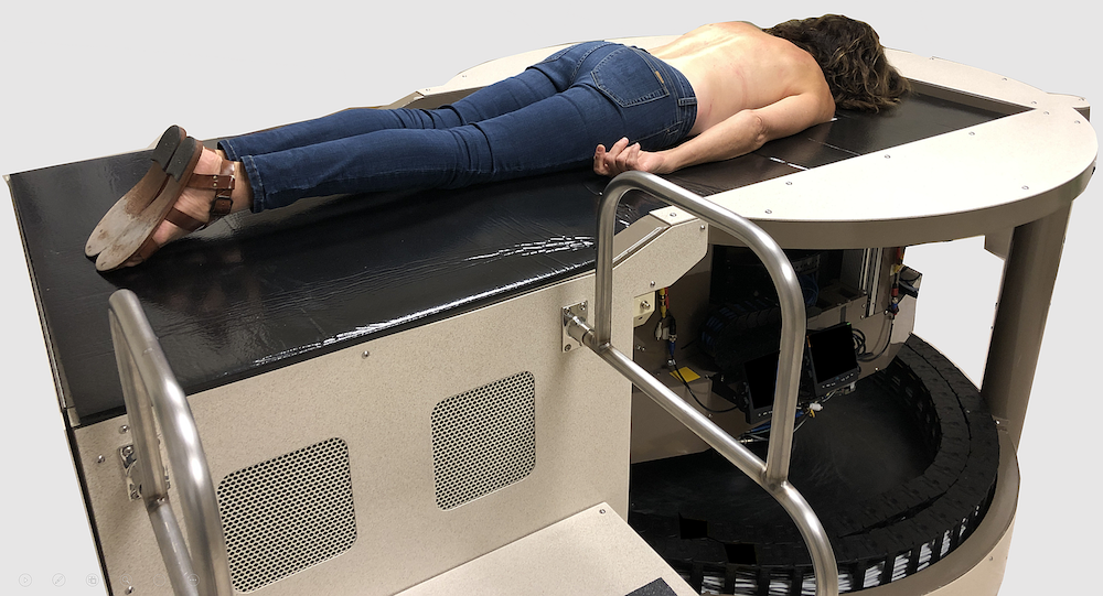 Figure 1: With dedicated breast CT, the woman lies prone on the table with the single breast to be imaged hanging through a hole in that table. There is no breast compression, and during a ten second breath-hold, the cone beam CT apparatus below the table scans the breast in one rotation providing all the necessary data to reconstruct the full 3D image data set from that breast.
The Boone lab at UC Davis has studied the potential of dedicated computed tomography for breast imaging for the past twenty years. Over this time, four breast CT systems have been designed, fabricated, and tested. Breast CT is a cone beam CT system in which the woman lies prone on the table, with the single breast to be imaged hanging pendant through that hole (Figure 1). The cone beam CT apparatus, primarily an x-ray tube and a flat panel detector, rotates around the breast and produces hundreds of low-dose projection images of the breast.
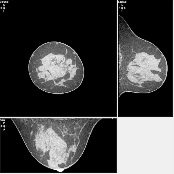 Figure 2: The reconstructed full data set from breast CT results in a high-resolution (150 µm voxels) depiction of the breast in three dimensions. Here, the coronal (upper left), sagittal (upper right), and axial (bottom) images of a breast are shown.
The projection images are reconstructed to produce a fully three-dimensional high-resolution image data set of the breast, as shown in Figure 2. It is worth noting that this is a true 3D depiction of the breast, something that tomosynthesis is not capable of producing due to its limited angle characteristics.
The breast CT systems in our laboratory have imaged almost 600 patients, using both non-contrast and (iodinated) contrast agent is injected into the arm vein. Because of the true three-dimensional nature of these image data sets, summation artifacts and other abnormalities associated with 2D mammography are overcome. Very importantly, by allowing the radiologist to view CT sections of the breast which eliminates the superposition of tissue, breast CT is far more robust to the dense breast than 2D mammography.
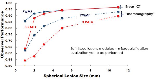 Figure 3: The results of observer performance studies comparing breast CT with digital mammography are shown, with the blue lines corresponding to the pre-whitened matched filter (a computer algorithm), and the red lines corresponding to the average performance of three dedicated breast imaging radiologists. Not only does breast CT outperform mammography, but the radiologists are also better to match the performance of the computer observer with breast CT, compared to mammography.
While the images (figure 2) are impressive, the performance of breast CT has been compared in observer performance studies using both computer observers and breast imaging radiologists, and these data are shown in figure 3. The computer observer (a pre-whitened matched filter) is known to outperform human observers in these types of studies, and this is seen in the figure. The computer observer performance significantly better with the breast CT image data set, then with the mammography data sets produced from the 3D CT volume. Another key observation in figure 3 is that the radiologists observers are better able to match the performance of the computer observer with breast CT image data, while their efficiency at replicating the performance of the computer observer with mammography data sets is significantly reduced.
The breast tomography project at UC Davis has produced dozens of peer-reviewed articles over the years, and has attracted over $20 million of external research funding. UC Davis patents on this technology have been licensed, and commercialization efforts are currently underway.
- AM Hernandez, CK Abbey, P Ghazi, GW Burkett, JM Boone, Effects of kV, filtration, dose and object size on soft tissue and iodine contrast in dedicated breast CT, Medical Physics 2020; 47:2896-2880
- F di Franco, A Sarno, G Mettivier, AM Hernandez, K Bliznakova, JM Boone, P Russo, GEOANT4 Monte Carlo simuilations for virtual clinical trials in breast x-ray imaging: Proof of concept, Physica Medica 2020; 74:133-142
- Daskalov S, Okkalidis N, Boone JM, et al. Anthropomorphic Physical Breast Phantom Based on Patient Breast CT Data: Preliminary Results. Mediterranean Conference on Medical and Biological Engineering and Computing; 2019: Springer, Cham: 367-374.
- Abbey CK, Bakic PR, Pokrajac DD, Maidment AD, Eckstein MP, Boone JM. Evaluation of non-Gaussian statistical properties in virtual breast phantoms. Journal of Medical Imaging 2019;6:025502.
- Aminololama-Shakeri S, Abbey CK, López JE, et al. Conspicuity of suspicious breast lesions on contrast enhanced breast CT compared to digital breast tomosynthesis and mammography. The British journal of radiology 2019;92:20181034.
- Ghazi P, Hernandez AM, Abbey C, Yang K, Boone JM. Shading artifact correction in breast CT using an interleaved deep learning segmentation and maximum likelihood polynomial fitting approach. Medical physics 2019.
- He Y, Liu Y, Dyer BA, et al. 3D-printed breast phantom for multi-purpose and multi-modality imaging. Quantitative imaging in medicine and surgery 2019;9:63.
- Hernandez AM, Becker AE, Boone JM. Updated breast CT dose coefficients (Dg NCT) using patient‐derived breast shapes and heterogeneous fibroglandular distributions. Medical physics 2019;46:1455-1466.
- Caballo M, Boone JM, Mann R, Sechopoulos I. An unsupervised automatic segmentation algorithm for breast tissue classification of dedicated breast computed tomography images. Medical physics. 2018;45(6):2542-2559.
- Lee J, Nishikawa RM, Reiser I, Boone JM. Neutrosophic segmentation of breast lesions for dedicated breast computed tomography. Journal of Medical Imaging. 2018;5(1):014505.
- Lee J, Nishikawa RM, Reiser I, Boone JM. Relationship between computer segmentation performance and computer classification performance in breast CT: A simulation study using RGI segmentation and LDA classification. Medical Physics. 2018;45(8):3650-3656.
- Sarno A, Mettivier G, Tucciariello RM, et al. Monte Carlo evaluation of glandular dose in cone-beam X-ray computed tomography dedicated to the breast: Homogeneous and heterogeneous breast models. Physica Medica. 2018;51:99-107.
- Abbey CK, Bakic PR, Pokrajac DD, Maidment AD, Eckstein MP, Boone JM. Evaluation of non-Gaussian statistical properties in virtual breast phantoms. Journal of Medical Imaging. 2019;6(2):025502.
- Lee, J, Nishikawa, RM, Reiser, I, Zuley, ML, Boone, JM. Lack of agreement between radiologists: implications for image-based model observers. Journal of Medical Imaging (Bellingham, Wash.), 4(2): 025502, 2017
- Lee, J, Nishikawa, RM, Reiser, I, Boone, JM. Optimal reconstruction and quantitative image features for computer-aided diagnosis tools for breast CT. Medical Physics, 44(5): 1846-1856, 2017
- Chaudhari, AJ, Ferrero, A, Godinez, F, Yang, K, Shelton, DK, Hunter, JC, Naguwa, SM, Boone, JM, Raychaudhuri, SP, Badawi, RD. High-resolution (18)F-FDG PET/CT for assessing disease activity in rheumatoid and psoriatic arthritis: findings of a prospective pilot study. The British journal of radiology, 89(1063): 20160138, 2016
- Gazi, PM, Aminololama-Shakeri, S, Yang, K, Boone, JM. Temporal subtraction contrast-enhanced dedicated breast CT. Physics in medicine and biology, 61(17): 6322-46, 2016
- Aminololama-Shakeri, Shadi; Abbey, Craig K; Gazi, Peymon; Prionas, Nicolas D; Nosratieh, Anita; Li, Chin-Shang; Boone, John M; Lindfors, Karen K; Differentiation of ductal carcinoma in-situ from benign micro-calcifications by dedicated breast computed tomography, European Journal of Radiology 85(1): 297-303; 2016
- Hernandez, AM; Seibert, JA; Boone, JM; Breast dose in mammography is about 30% lower when realistic heterogeneous glandular distributions are considered, Med Phys 42(11): 6337-6348; 2015 [Editor’s Pick for 2015, and Best Paper of 2015]
- Nosratieh, Anita; Hernandez, Andrew; Shen, Sam Z; Yaffe, Martin J; Seibert, J Anthony; Boone, JM, Mean glandular dose coefficients (DgN) for x-ray spectra used in contemporary breast imaging systems, PMB 60(18): 7179; 20125
- Lee, Juhun; Nishikawa, Robert M; Reiser, Ingrid; Boone, John M; Lindfors, Karen K; Local curvature analysis for classifying breast tumors: Preliminary analysis in dedicated breast CT, Medical Physics 42(9):5479-5489; 2015
- Kuo, Hsien-Chi; Giger, Maryellen L; Reiser, Ingrid; Drukker, Karen; Boone, John M; Lindfors, Karen K; Yang, Kai; Edwards, Alexandra; Impact of lesion segmentation metrics on computer-aided diagnosis/detection in breast computed tomography, Journal of Medical Imaging:1(3):031012-031012: 2014
- Gazi, Peymon M; Yang, Kai; Burkett Jr, George W; Aminololama-Shakeri, Shadi; Seibert, J Anthony; Boone, John M; Evolution of spatial resolution in breast CT at UC Davis. Med Phys 42(4): 1973-1981: 2015
- Chen, L; Boone, JM; Abbey, CK; Hargreaves, J; Bateni, C; Lindfors, KK; Yang, K; Nosratieh, A; Hernandez, A; Gazi, P; Simulated lesion, human observer performance comparison between thin-section dedicated breast CT images versus computed thick-section simulated projection images of the breast, Physics in medicine and biology 60(8): 3347: 2015
- Prionas, ND; Aminololama-Shakeri, S; Yang, Ki; Martinez, SR; Lindfors, KK; Boone, JM, Contrast-enhanced dedicated breast CT detection of invasive breast cancer preceding mammographic diagnosis, Radiology Case Reports 10; 2015
- Yang, Kai; Burkett Jr, George; Boone, John M; A breast-specific, negligible-dose scatter correction technique for dedicated cone-beam breast CT: a physics-based approach to improve Hounsfield Unit accuracy, PMB 59(21); 6487: 2014
- Santos J, Chaudhari AJ, Joshi AA, Ferrero A, Yang K, Boone JM, et al. Non-rigid registration of serial dedicated breast CT, longitudinal dedicated breast CT and PET/CT images using the diffeomorphic demons method. Physica Medica. 2014.
- Kuo H-C, Giger ML, Reiser I, Drukker K, Boone JM, Lindfors KK, et al. Segmentation of breast masses on dedicated breast computed tomography and three-dimensional breast ultrasound images. Journal of Medical Imaging. 2014;1(1):014501-.
- Kuo H-C, Giger ML, Reiser I, Boone JM, Lindfors KK, Yang K, et al. Level set segmentation of breast masses in contrast-enhanced dedicated breast CT and evaluation of stopping criteria. Journal of digital imaging. 2014;27(2):237-47.
- Bian J, Yang K, Boone JM, Han X, Sidky EY, Pan X. Investigation of iterative image reconstruction in low-dose breast CT. Physics in Medicine and Biology. 2014;59(11):2659.
- Abbey CK, Gallas BD, Boone JM, Niklason LT, Hadjiiski LM, Sahiner B, et al. Comparative Statistical Properties of Expected Utility and Area Under the ROC Curve for Laboratory Studies of Observer Performance in Screening Mammography. Academic Radiology. 2014;21(4):481-90.
- Chen L, Abbey CK, Boone JM. Association between power law coefficients of the anatomical noise power spectrum and lesion detectability in breast imaging modalities. Physics in Medicine and Biology. 2013;58(6):1663.
- Abbey CK, Eckstein MP, Boone JM. Estimating the relative utility of screening mammography. Medical Decision Making. 2013;33(4):510-20.
- Reiser I, Nishikawa R, Giger M, Boone J, Lindfors K, Yang K. Automated detection of mass lesions in dedicated breast CT: A preliminary study. Medical Physics. 2012;39(2):866-73.
- Prionas ND, McKenney SE, Stern RL, Boone JM. Kilovoltage rotational external beam radiotherapy on a breast computed tomography platform: a feasibility study. International Journal of Radiation Oncology Biology Physics. 2012;84(2):533-9.
- Prionas ND, Burkett GW, McKenney SE, Chen L, Stern RL, Boone JM. Development of a patient-specific two-compartment anthropomorphic breast phantom. Physics in Medicine and Biology. 2012;57(13):4293.
- Packard NJ, Abbey CK, Yang K, Boone JM. Effect of slice thickness on detectability in breast CT using a pre-whitened matched filter and simulated mass lesions. Medical Physics. 2012;39(4):1818-30.
- Nosratieh A, Yang K, Aminololama-Shakeri S, Boone JM. Comprehensive assessment of the slice sensitivity profiles in breast tomosynthesis and breast CT. Medical Physics. 2012;39(12):7254-61.
- Kalender WA, Beister M, Boone JM, Kolditz D, Vollmar SV, Weigel MC. High-resolution spiral CT of the breast at very low dose: concept and feasibility considerations. European Radiology. 2012;22(1):1-8.
- Huang S-Y, Yang K, Abbey CK, Boone JM. A semiempirical linear model of indirect, flat-panel x-ray detectors. Medical Physics. 2012;39(4):2108-18.
- Chen L, Abbey CK, Nosratieh A, Lindfors KK, Boone JM. Anatomical complexity in breast parenchyma and its implications for optimal breast imaging strategies. Medical Physics. 2012;39(3):1435-41.
- Abbey CK, Nosratieh A, Sohl-Dickstein J, Yang K, Boone JM. Non-Gaussian statistical properties of breast images. Medical Physics. 2012;39(11):7121-30.
- Prionas ND, Huang S-Y, Boone JM. Experimentally determined spectral optimization for dedicated breast computed tomography. Medical Physics. 2011;38(2):646-55.
- Huang S-Y, Boone JM, Yang K, Packard NJ, McKenney SE, Prionas ND, et al. The characterization of breast anatomical metrics using dedicated breast CT. Medical Physics. 2011;38(4):2180-91.
- Yang K, Huang S-Y, Packard NJ, Boone JM. Noise variance analysis using a flat panel x-ray detector: A method for additive noise assessment with application to breast CT applications. Medical Physics. 2010;37(7):3527-37.
- Prionas ND, Lindfors KK, Ray S, Huang S-Y, Beckett LA, Monsky WL, et al. Contrast-enhanced Dedicated Breast CT: Initial Clinical Experience 1. Radiology. 2010;256(3):714-23.
- Lindfors KK, Boone JM, Newell MS, D'Orsi CJ. Dedicated breast computed tomography: the optimal cross-sectional imaging solution? Radiologic Clinics of North America. 2010;48(5):1043-54.
- Boone JM, Yang K, Burkett GW, Packard NJ, Huang S-Y, Bowen S, et al. An x-ray computed tomography/positron emission tomography system designed specifically for breast imaging. Technology in Cancer Research & Treatment. 2010;9(1):29.
- Alonzo-Proulx O, Packard N, Boone J, Al-Mayah A, Brock K, Shen S, et al. Validation of a method for measuring the volumetric breast density from digital mammograms. Physics in Medicine and Biology. 2010;55(11):3027.