Tumor microbiome linked to immunotherapy success in sarcoma patients
New UC Davis study finds relationship between tumor microbiome and immune system in patients with soft tissue sarcoma
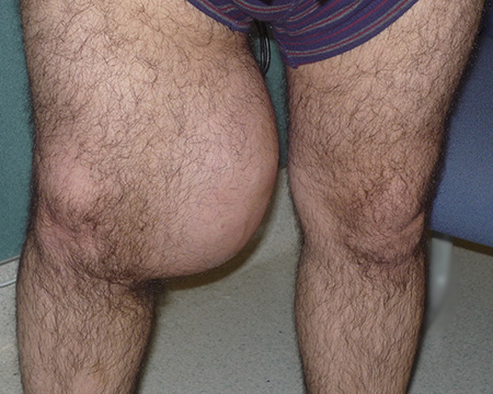
In a significant new study, UC Davis Comprehensive Cancer Center researchers have uncovered a link between a patient’s microbiome and their immune system that can potentially be used to improve the treatment of soft tissue sarcoma. This type of cancer is found in connective tissues like muscle, fat and nerves.
Findings from the study were published in the Journal for Immunotherapy of Cancer.
“The study’s data show new lines of research in the paradigm-shifting concept that the microbiome of a patient and their immune system can interact and shape one another, as well as be potentially engineered to improve patient outcomes,” said Robert Canter, the lead author of the study and chief of the Division of Surgical Oncology.
The gut microbiome is made of microorganisms in the digestive tract that include bacteria, fungi and viruses. Microbial communities have also been found in other parts of the body, including the mouth, lungs and skin. And now the study shows they are also found in tumor cells.
“We found that soft tissue sarcomas harbor a quantifiable amount of microbiome within the tumor environment. Most importantly, we found that the amount of microbiome at diagnosis may be linked with the patient’s prognosis,” Canter added.
Although the levels of microbes are low, the study findings are significant because many tumors, especially sarcomas, were believed to be sterile.
We found that soft tissue sarcomas harbor a quantifiable amount of microbiome within the tumor environment. Most importantly, we found that the amount of microbiome at diagnosis may be linked with the patient’s prognosis.”—Robert Canter, chief, UC Davis Division of Surgical Oncology
Viruses within the microbiome may attract cancer-fighting cells
The UC Davis researchers also uncovered how the microbiome within a sarcoma tumor plays a role in attracting specific types of immune cells like cancer-fighting natural killer cells. Canter said that’s important because the higher the rate of natural killer cell infiltration in a tumor, the greater the chance that the sarcoma won’t spread to other parts of the body. Natural killer cells are a prime target for improving the effectiveness of immunotherapy.
The team found that viruses within the microbiome of a tumor appear to impact the amount of natural killer cells found in sarcomas and, for that reason, affect survival rates. Specifically, the study found a strong positive correlation between the presence of Respirovirus, a genre of viruses known for causing respiratory illnesses, and the presence of natural killer cells in the tumor. Canter and his colleagues are now considering ways to create viruses to attract more cancer-killing immune cells.
“It has become clear that the microbiome in the gut and other parts of the body has a major impact on human health and disease. Amazingly, it shapes the immune system throughout the body and, because of its interaction with the immune system, we now know it also has a big role in how the body responds to cancer and cancer treatments like immunotherapy,” Canter said.
Cross-campus collaboration
The authors obtained tumor and stool samples from 15 adult patients with non-metastatic soft tissue sarcoma, which was studied for a median of 24 months. Analysis revealed that most of the tumors were advanced stage III (87%) and affected a patient’s limb (67%).
Tissue samples were sent to the university’s Genome Center in Davis for sequencing and to the Flow Cytometry Shared Resource Laboratory on the UC Davis medical campus in Sacramento for immune profiling. Patients were monitored for two years as part of their cancer treatment follow-up.
Canter said past research has shown the existence of microbiome inside tumors across several cancer types, including breast, lung, pancreas and melanoma. For that reason, he said further research into the connection between microbiome and the immune system in other cancer types is warranted.
Acknowledgments
Coauthors on the study are Lauren Perry, Sylvia Cruz, Kara Kleber, Sean Judge, Morgan Darrow, Louis Jones, Ugur Basmaci, Nikhil Joshi, Matthew Settles, Blythe Durbin-Johnson, Alicia Gingrich, Arta Monjazeb, Janai Carr-Ascher, Steven Thorpe, William Murphy, and Jonathan Eisen.
This work was supported by the UC Davis Comprehensive Cancer Center and the UC Davis Flow Cytometry Shared Resource Laboratory, with funding from National Cancer Institute grants P30 CA093373, S10 OD018223 and S10 RR 026825. Specimens were provided by the UC Davis Pathology Biorepository, funded by the UC Davis Comprehensive Cancer and the UC Davis Department of Pathology and Laboratory Medicine. The sequencing was carried out at the DNA Technologies and Expression Analysis Cores at the UC Davis Genome Center, supported by National Institutes of Health (NIH) Shared Instrumentation Grant 1S10OD010786-01.
UC Davis Comprehensive Cancer Center
UC Davis Comprehensive Cancer Center is the only National Cancer Institute-designated center serving the Central Valley and inland Northern California, a region of more than 6 million people. Its specialists provide compassionate, comprehensive care for more than 100,000 adults and children every year and access to more than 200 active clinical trials at any given time. Its innovative research program engages more than 240 scientists at UC Davis who work collaboratively to advance discovery of new tools to diagnose and treat cancer. Patients have access to leading-edge care, including immunotherapy and other targeted treatments. Its Office of Community Outreach and Engagement addresses disparities in cancer outcomes across diverse populations, and the cancer center provides comprehensive education and workforce development programs for the next generation of clinicians and scientists. For more information, visit cancer.ucdavis.edu.


